

passive absorption, multivalent chelation, or covalent bond formation as
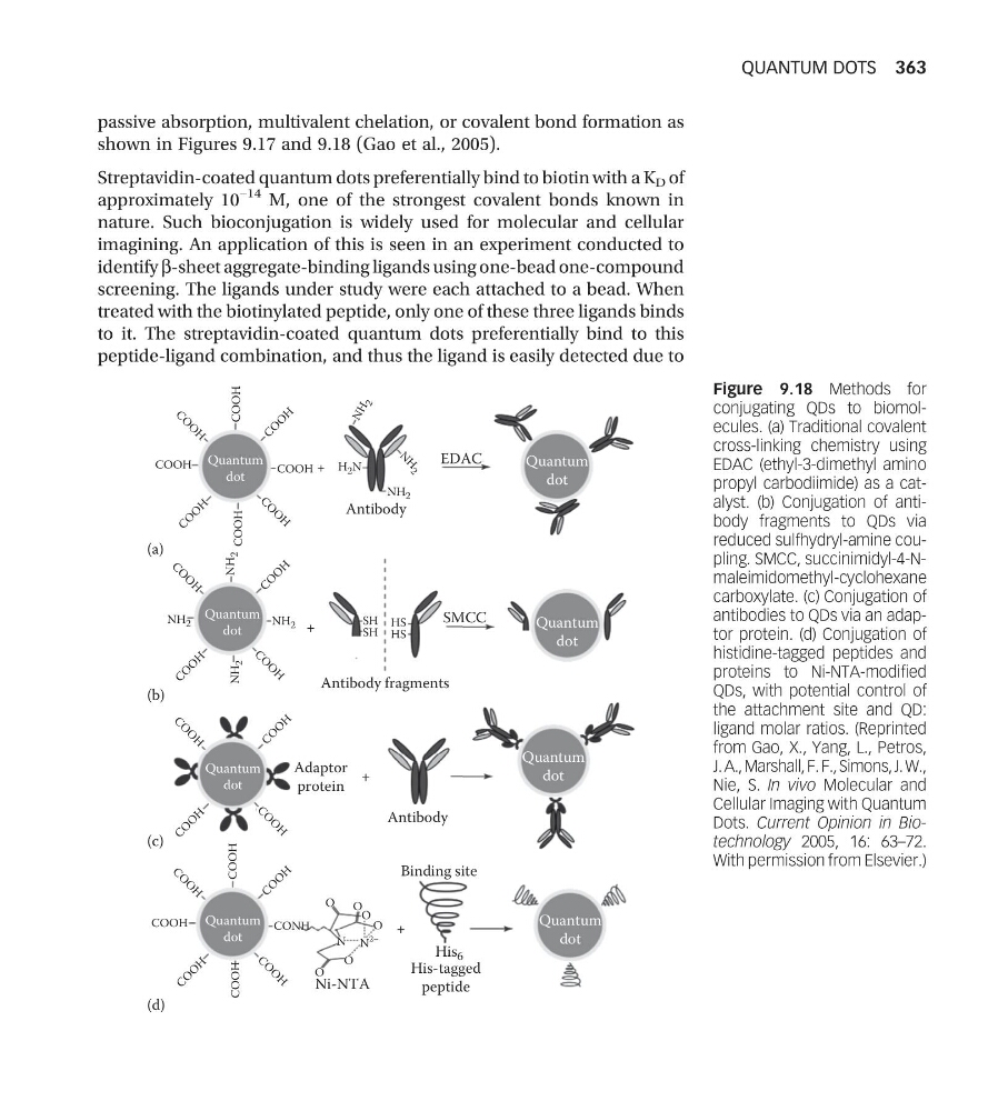
shown in Figures 9.17 and 9.18 (Gao et al., 2005).
Streptavidin-coated quantum dots preferentially bind to biotin with a K D of
approximately 10
−14 M, one of the strongest covalent bonds known in
nature. Such bioconjugation is widely used for molecular and cellular
imagining. An application of this is seen in an experiment conducted to
identify b-sheet aggregate-binding ligands using one-bead one-compound
screening. The ligands under study were each attached to a bead. When
treated with the biotinylated peptide, only one of these three ligands binds
to it. The streptavidin-coated quantum dots preferentially bind to this
peptide-ligand combination, and thus the ligand is easily detected due to
Quantum
dot
Quantum
dot
Quantum
dot
Quantum
dot
Quantum
dot
Quantum
dot
Antibody fragments
Adaptor
protein
Antibody
Binding site
His 6
Ni-NTA
His-tagged
peptide
Quantum
dot
Quantum
dot
Antibody
+
+
+
+
C O
O
H
C O
O
H
COOH
COOH
C
O
O
H
C
O
O
H
COOH
H 2 N
NH 2
NH 2
NH
2
NH
2
N
H
2
N H
2
NH 2
COOH
C O
O
H
C O
O
H
C
O
O
H
C O
O
H
COOH
COOH
C
O
O
H
CONH
O
O
O
O
O
O
N N
2–
COOH
C O
O
H
C
O
O
H
C
O
O
H
C O
O
H
C
O
O
H
C O
O
H
C
O
O
H
EDAC
SMCC
HS
HS
SH
SH
(a)
(b)
(c)
(d)
Figure 9.18 Methods for
conjugating QDs to biomolecules. (a) Traditional covalent
cross-linking chemistry using
EDAC (ethyl-3-dimethyl amino
propyl carbodiimide) as a catalyst. (b) Conjugation of antibody fragments to QDs via
reduced sulfhydryl-amine coupling. SMCC, succinimidyl-4-Nmaleimidomethyl-cyclohexane
carboxylate. (c) Conjugation of
antibodies to QDs via an adaptor protein. (d) Conjugation of
histidine-tagged peptides and
proteins to Ni-NTA-modified
QDs, with potential control of
the attachment site and QD:
ligand molar ratios. (Reprinted
from Gao, X., Yang, L., Petros,
J. A., Marshall, F. F., Simons, J. W.,
Nie, S. In vivo Molecular and
Cellular Imaging with Quantum
Dots. Current Opinion in Biotechnology 2005, 16: 63–72.
With permission from Elsevier.)
QUANTUM DOTS 363
shown in Figures 9.17 and 9.18 (Gao et al., 2005).
Streptavidin-coated quantum dots preferentially bind to biotin with a K D of
approximately 10
−14 M, one of the strongest covalent bonds known in
nature. Such bioconjugation is widely used for molecular and cellular
imagining. An application of this is seen in an experiment conducted to
identify b-sheet aggregate-binding ligands using one-bead one-compound
screening. The ligands under study were each attached to a bead. When
treated with the biotinylated peptide, only one of these three ligands binds
to it. The streptavidin-coated quantum dots preferentially bind to this
peptide-ligand combination, and thus the ligand is easily detected due to
Quantum
dot
Quantum
dot
Quantum
dot
Quantum
dot
Quantum
dot
Quantum
dot
Antibody fragments
Adaptor
protein
Antibody
Binding site
His 6
Ni-NTA
His-tagged
peptide
Quantum
dot
Quantum
dot
Antibody
+
+
+
+
C O
O
H
C O
O
H
COOH
COOH
C
O
O
H
C
O
O
H
COOH
H 2 N
NH 2
NH 2
NH
2
NH
2
N
H
2
N H
2
NH 2
COOH
C O
O
H
C O
O
H
C
O
O
H
C O
O
H
COOH
COOH
C
O
O
H
CONH
O
O
O
O
O
O
N N
2–
COOH
C O
O
H
C
O
O
H
C
O
O
H
C O
O
H
C
O
O
H
C O
O
H
C
O
O
H
EDAC
SMCC
HS
HS
SH
SH
(a)
(b)
(c)
(d)
Figure 9.18 Methods for
conjugating QDs to biomolecules. (a) Traditional covalent
cross-linking chemistry using
EDAC (ethyl-3-dimethyl amino
propyl carbodiimide) as a catalyst. (b) Conjugation of antibody fragments to QDs via
reduced sulfhydryl-amine coupling. SMCC, succinimidyl-4-Nmaleimidomethyl-cyclohexane
carboxylate. (c) Conjugation of
antibodies to QDs via an adaptor protein. (d) Conjugation of
histidine-tagged peptides and
proteins to Ni-NTA-modified
QDs, with potential control of
the attachment site and QD:
ligand molar ratios. (Reprinted
from Gao, X., Yang, L., Petros,
J. A., Marshall, F. F., Simons, J. W.,
Nie, S. In vivo Molecular and
Cellular Imaging with Quantum
Dots. Current Opinion in Biotechnology 2005, 16: 63–72.
With permission from Elsevier.)
QUANTUM DOTS 363