

and their coercivity in magnetic materials. Properties of an ensemble of
quantum dots are additionally determined by particle size distribution
and differences in morphology within the ensemble.
9.4.1 Optical properties of quantum dots
Quantum confinement gives quantum dots distinct optical properties compared to bulk semiconductors. Quantum confinement occurs when the
radius of the quantum dot is smaller than the exciton radius in the corresponding bulk semiconductor. For CdSe, this transition occurs at a radius of
5.6 nm (Nirmal, 1999). Below this size, the quantum dot has clearly distinguishable discrete energy levels, and its band gap is shifted by an energy
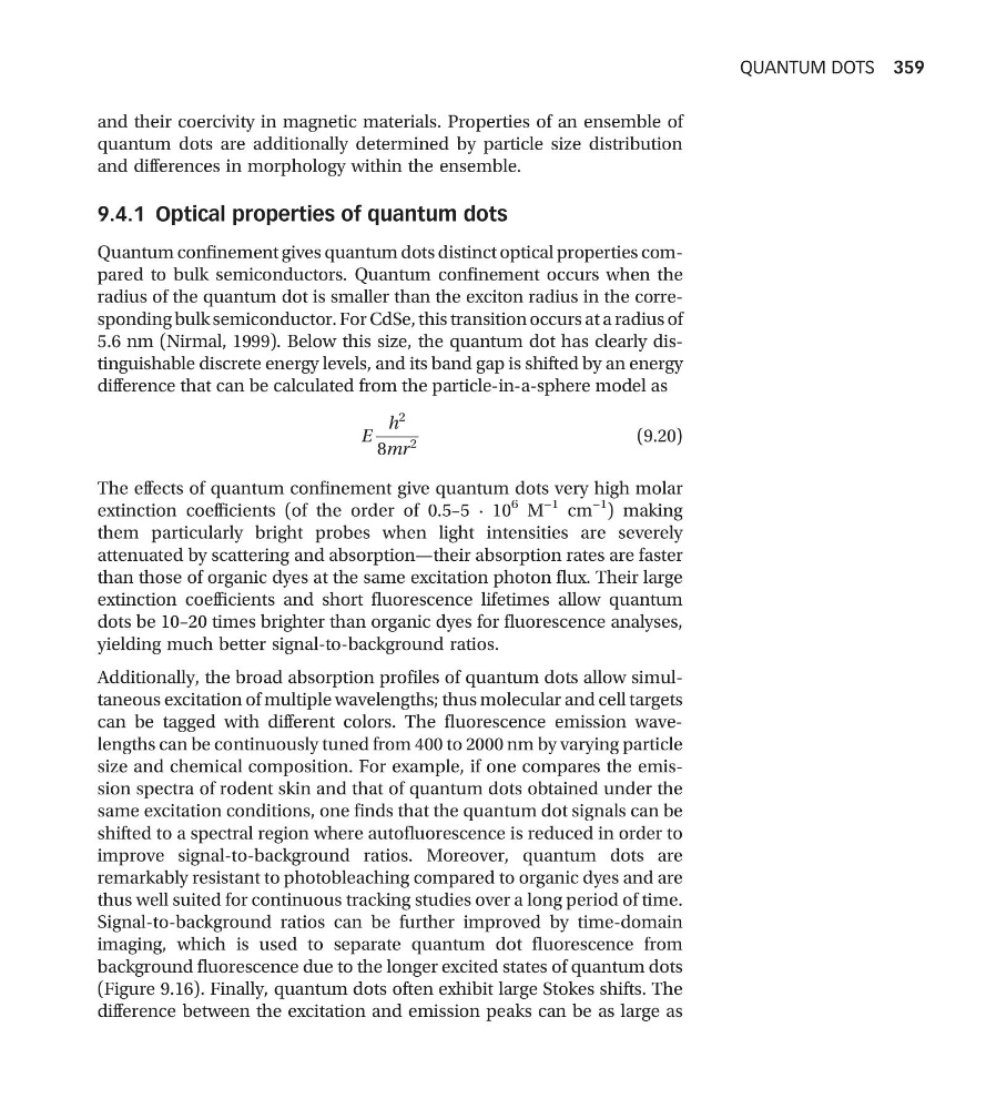
difference that can be calculated from the particle-in-a-sphere model as
E
h
2
8mr
2
(9.20)
The effects of quantum confinement give quantum dots very high molar
extinction coefficients (of the order of 0.5–5 · 10
6 M
−1 cm
−1 ) making
them particularly bright probes when light intensities are severely
attenuated by scattering and absorption—their absorption rates are faster
than those of organic dyes at the same excitation photon flux. Their large
extinction coefficients and short fluorescence lifetimes allow quantum
dots be 10–20 times brighter than organic dyes for fluorescence analyses,
yielding much better signal-to-background ratios.
Additionally, the broad absorption profiles of quantum dots allow simultaneous excitation of multiple wavelengths; thus molecular and cell targets
can be tagged with different colors. The fluorescence emission wavelengths can be continuously tuned from 400 to 2000 nm by varying particle
size and chemical composition. For example, if one compares the emission spectra of rodent skin and that of quantum dots obtained under the
same excitation conditions, one finds that the quantum dot signals can be
shifted to a spectral region where autofluorescence is reduced in order to
improve signal-to-background ratios. Moreover, quantum dots are
remarkably resistant to photobleaching compared to organic dyes and are
thus well suited for continuous tracking studies over a long period of time.
Signal-to-background ratios can be further improved by time-domain
imaging, which is used to separate quantum dot fluorescence from
background fluorescence due to the longer excited states of quantum dots
(Figure 9.16). Finally, quantum dots often exhibit large Stokes shifts. The
difference between the excitation and emission peaks can be as large as
QUANTUM DOTS 359
quantum dots are additionally determined by particle size distribution
and differences in morphology within the ensemble.
9.4.1 Optical properties of quantum dots
Quantum confinement gives quantum dots distinct optical properties compared to bulk semiconductors. Quantum confinement occurs when the
radius of the quantum dot is smaller than the exciton radius in the corresponding bulk semiconductor. For CdSe, this transition occurs at a radius of
5.6 nm (Nirmal, 1999). Below this size, the quantum dot has clearly distinguishable discrete energy levels, and its band gap is shifted by an energy
difference that can be calculated from the particle-in-a-sphere model as
E
h
2
8mr
2
(9.20)
The effects of quantum confinement give quantum dots very high molar
extinction coefficients (of the order of 0.5–5 · 10
6 M
−1 cm
−1 ) making
them particularly bright probes when light intensities are severely
attenuated by scattering and absorption—their absorption rates are faster
than those of organic dyes at the same excitation photon flux. Their large
extinction coefficients and short fluorescence lifetimes allow quantum
dots be 10–20 times brighter than organic dyes for fluorescence analyses,
yielding much better signal-to-background ratios.
Additionally, the broad absorption profiles of quantum dots allow simultaneous excitation of multiple wavelengths; thus molecular and cell targets
can be tagged with different colors. The fluorescence emission wavelengths can be continuously tuned from 400 to 2000 nm by varying particle
size and chemical composition. For example, if one compares the emission spectra of rodent skin and that of quantum dots obtained under the
same excitation conditions, one finds that the quantum dot signals can be
shifted to a spectral region where autofluorescence is reduced in order to
improve signal-to-background ratios. Moreover, quantum dots are
remarkably resistant to photobleaching compared to organic dyes and are
thus well suited for continuous tracking studies over a long period of time.
Signal-to-background ratios can be further improved by time-domain
imaging, which is used to separate quantum dot fluorescence from
background fluorescence due to the longer excited states of quantum dots
(Figure 9.16). Finally, quantum dots often exhibit large Stokes shifts. The
difference between the excitation and emission peaks can be as large as
QUANTUM DOTS 359