

The most basic method by which photon energy can be harnessed by
molecular systems is through the exploitation of the specific absorptionfluorescence characteristics of dye molecules. Let us examine the concept
of energy transport between dyes using a model system of two interacting
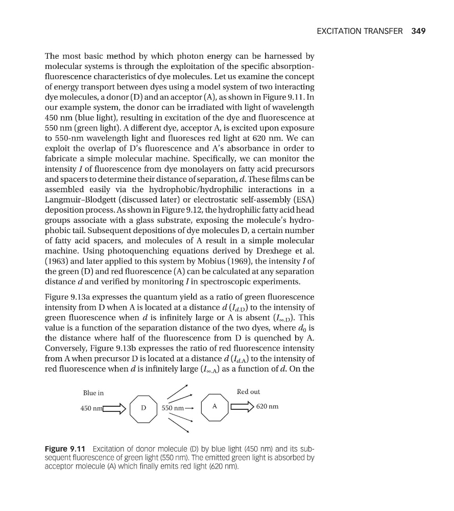
dye molecules, a donor (D) and an acceptor (A), as shown in Figure 9.11. In
our example system, the donor can be irradiated with light of wavelength
450 nm (blue light), resulting in excitation of the dye and fluorescence at
550 nm (green light). A different dye, acceptor A, is excited upon exposure
to 550-nm wavelength light and fluoresces red light at 620 nm. We can
exploit the overlap of D’s fluorescence and A’s absorbance in order to
fabricate a simple molecular machine. Specifically, we can monitor the
intensity I of fluorescence from dye monolayers on fatty acid precursors
and spacers to determine their distance of separation, d. These films can be
assembled easily via the hydrophobic/hydrophilic interactions in a
Langmuir–Blodgett (discussed later) or electrostatic self-assembly (ESA)
deposition process. As shown in Figure 9.12, the hydrophilic fatty acid head
groups associate with a glass substrate, exposing the molecule’s hydrophobic tail. Subsequent depositions of dye molecules D, a certain number
of fatty acid spacers, and molecules of A result in a simple molecular
machine. Using photoquenching equations derived by Drexhege et al.
(1963) and later applied to this system by Mobius (1969), the intensity I of
the green (D) and red fluorescence (A) can be calculated at any separation
distance d and verified by monitoring I in spectroscopic experiments.
Figure 9.13a expresses the quantum yield as a ratio of green fluorescence
intensity from D when A is located at a distance d (I d.D ) to the intensity of
green fluorescence when d is infinitely large or A is absent (I ∞.D ). This
value is a function of the separation distance of the two dyes, where d 0 is
the distance where half of the fluorescence from D is quenched by A.
Conversely, Figure 9.13b expresses the ratio of red fluorescence intensity
from A when precursor D is located at a distance d (I d.A ) to the intensity of
red fluorescence when d is infinitely large (I ∞.A ) as a function of d. On the
Blue in
Red out
D
450 nm
A
550 nm
620 nm
Figure 9.11 Excitation of donor molecule (D) by blue light (450 nm) and its subsequent fluorescence of green light (550 nm). The emitted green light is absorbed by
acceptor molecule (A) which finally emits red light (620 nm).
EXCITATION TRANSFER 349
molecular systems is through the exploitation of the specific absorptionfluorescence characteristics of dye molecules. Let us examine the concept
of energy transport between dyes using a model system of two interacting
dye molecules, a donor (D) and an acceptor (A), as shown in Figure 9.11. In
our example system, the donor can be irradiated with light of wavelength
450 nm (blue light), resulting in excitation of the dye and fluorescence at
550 nm (green light). A different dye, acceptor A, is excited upon exposure
to 550-nm wavelength light and fluoresces red light at 620 nm. We can
exploit the overlap of D’s fluorescence and A’s absorbance in order to
fabricate a simple molecular machine. Specifically, we can monitor the
intensity I of fluorescence from dye monolayers on fatty acid precursors
and spacers to determine their distance of separation, d. These films can be
assembled easily via the hydrophobic/hydrophilic interactions in a
Langmuir–Blodgett (discussed later) or electrostatic self-assembly (ESA)
deposition process. As shown in Figure 9.12, the hydrophilic fatty acid head
groups associate with a glass substrate, exposing the molecule’s hydrophobic tail. Subsequent depositions of dye molecules D, a certain number
of fatty acid spacers, and molecules of A result in a simple molecular
machine. Using photoquenching equations derived by Drexhege et al.
(1963) and later applied to this system by Mobius (1969), the intensity I of
the green (D) and red fluorescence (A) can be calculated at any separation
distance d and verified by monitoring I in spectroscopic experiments.
Figure 9.13a expresses the quantum yield as a ratio of green fluorescence
intensity from D when A is located at a distance d (I d.D ) to the intensity of
green fluorescence when d is infinitely large or A is absent (I ∞.D ). This
value is a function of the separation distance of the two dyes, where d 0 is
the distance where half of the fluorescence from D is quenched by A.
Conversely, Figure 9.13b expresses the ratio of red fluorescence intensity
from A when precursor D is located at a distance d (I d.A ) to the intensity of
red fluorescence when d is infinitely large (I ∞.A ) as a function of d. On the
Blue in
Red out
D
450 nm
A
550 nm
620 nm
Figure 9.11 Excitation of donor molecule (D) by blue light (450 nm) and its subsequent fluorescence of green light (550 nm). The emitted green light is absorbed by
acceptor molecule (A) which finally emits red light (620 nm).
EXCITATION TRANSFER 349