

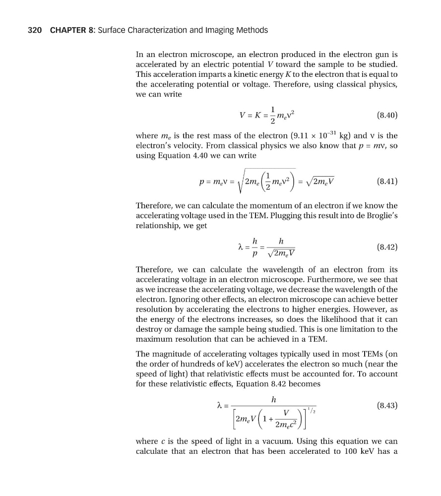
In an electron microscope, an electron produced in the electron gun is
accelerated by an electric potential V toward the sample to be studied.
This acceleration imparts a kinetic energy K to the electron that is equal to
the accelerating potential or voltage. Therefore, using classical physics,
we can write
V = K =
1
2
m e n
2
(8.40)
where m e is the rest mass of the electron (9.11 × 10
–31 kg) and n is the
electron’s velocity. From classical physics we also know that p = mn, so
using Equation 4.40 we can write
p = m e n =
ffiffiffiffiffiffiffiffiffiffiffiffiffiffiffiffiffiffiffiffiffiffiffiffiffiffiffiffiffi
2m e
1
2
m e n
2
s
=
ffiffiffiffiffiffiffiffiffiffiffiffi
2m e V
p
(8.41)
Therefore, we can calculate the momentum of an electron if we know the
accelerating voltage used in the TEM. Plugging this result into de Broglie’s
relationship, we get
l =
h
p
=
h
ffiffiffiffiffiffiffiffiffiffiffiffi
2m e V
p
(8.42)
Therefore, we can calculate the wavelength of an electron from its
accelerating voltage in an electron microscope. Furthermore, we see that
as we increase the accelerating voltage, we decrease the wavelength of the
electron. Ignoring other effects, an electron microscope can achieve better
resolution by accelerating the electrons to higher energies. However, as
the energy of the electrons increases, so does the likelihood that it can
destroy or damage the sample being studied. This is one limitation to the
maximum resolution that can be achieved in a TEM.
The magnitude of accelerating voltages typically used in most TEMs (on
the order of hundreds of keV) accelerates the electron so much (near the
speed of light) that relativistic effects must be accounted for. To account
for these relativistic effects, Equation 8.42 becomes
l =
h
2m e V 1 +
V
2m e c 2
! 1
2
=
(8.43)
where c is the speed of light in a vacuum. Using this equation we can
calculate that an electron that has been accelerated to 100 keV has a
CHAPTER 8: Surface Characterization and Imaging Methods
320
accelerated by an electric potential V toward the sample to be studied.
This acceleration imparts a kinetic energy K to the electron that is equal to
the accelerating potential or voltage. Therefore, using classical physics,
we can write
V = K =
1
2
m e n
2
(8.40)
where m e is the rest mass of the electron (9.11 × 10
–31 kg) and n is the
electron’s velocity. From classical physics we also know that p = mn, so
using Equation 4.40 we can write
p = m e n =
ffiffiffiffiffiffiffiffiffiffiffiffiffiffiffiffiffiffiffiffiffiffiffiffiffiffiffiffiffi
2m e
1
2
m e n
2
s
=
ffiffiffiffiffiffiffiffiffiffiffiffi
2m e V
p
(8.41)
Therefore, we can calculate the momentum of an electron if we know the
accelerating voltage used in the TEM. Plugging this result into de Broglie’s
relationship, we get
l =
h
p
=
h
ffiffiffiffiffiffiffiffiffiffiffiffi
2m e V
p
(8.42)
Therefore, we can calculate the wavelength of an electron from its
accelerating voltage in an electron microscope. Furthermore, we see that
as we increase the accelerating voltage, we decrease the wavelength of the
electron. Ignoring other effects, an electron microscope can achieve better
resolution by accelerating the electrons to higher energies. However, as
the energy of the electrons increases, so does the likelihood that it can
destroy or damage the sample being studied. This is one limitation to the
maximum resolution that can be achieved in a TEM.
The magnitude of accelerating voltages typically used in most TEMs (on
the order of hundreds of keV) accelerates the electron so much (near the
speed of light) that relativistic effects must be accounted for. To account
for these relativistic effects, Equation 8.42 becomes
l =
h
2m e V 1 +
V
2m e c 2
! 1
2
=
(8.43)
where c is the speed of light in a vacuum. Using this equation we can
calculate that an electron that has been accelerated to 100 keV has a
CHAPTER 8: Surface Characterization and Imaging Methods
320