

relativistic wavelength of ~3.7 pm. For a TEM, we can approximate the
maximum resolution by modifying Equation 8.38 to
d ≈ 0:61
l
b
(8.44)
where b in this case is a property of the electron “lens” used to focus the
electron beam. Therefore, using Equation 8.44 we see that with a 100-keV
electron beam (l ~ 4 pm), we can achieve a theoretical maximum resolution (d ~ several picometers) that is smaller than the diameter of an
atom. While such maximum resolution is not feasible because we are
unable to build perfect electron lenses, it highlights the fact that TEM is
able to achieve a high level of resolution.
8.7.3.2 TEM instrumentation
In a simplified way, a transmission electron microscope operates like a
slide projector. In a slide projector, a beam of light is transmitted through
a slide. Some of the light is reflected or absorbed by the slide, so when the
transmitted light is projected onto a screen, an image is produced. A TEM
functions in essentially the same manner, with the only difference being
that a beam of electrons is transmitted through the sample rather than a
beam of visible light.
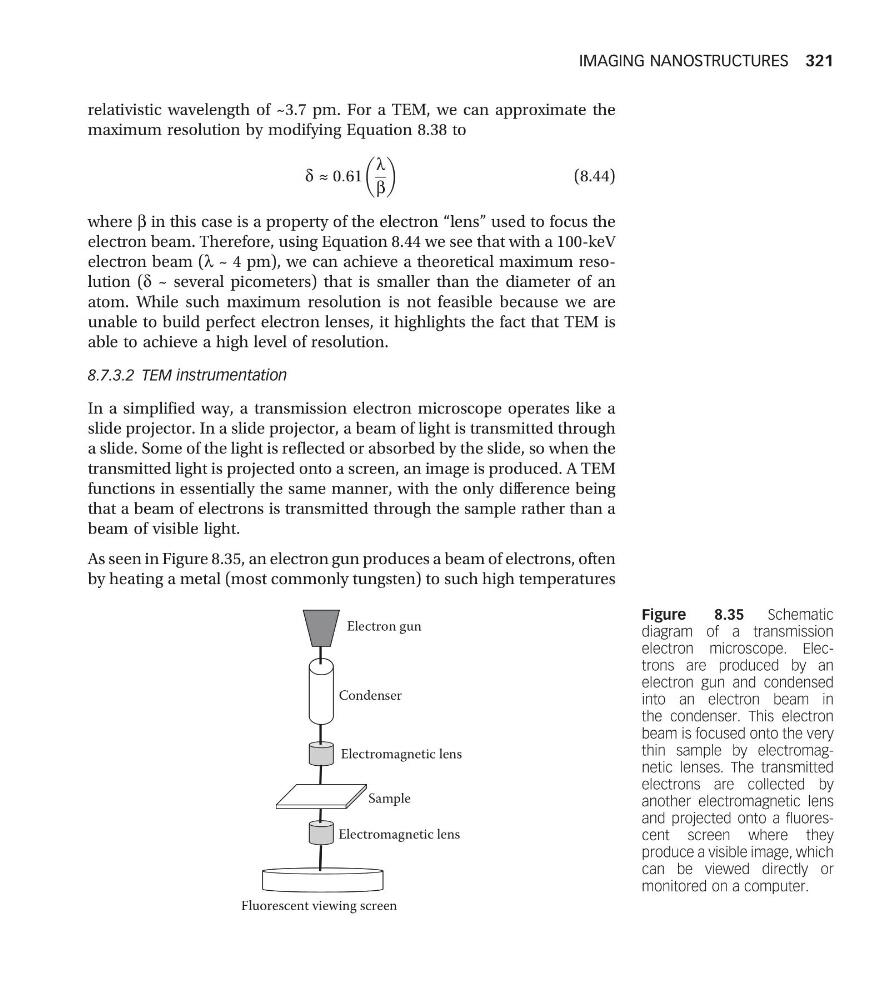
As seen in Figure 8.35, an electron gun produces a beam of electrons, often
by heating a metal (most commonly tungsten) to such high temperatures
Electron gun
Condenser
Electromagnetic lens
Sample
Electromagnetic lens
Fluorescent viewing screen
Figure
8.35
Schematic
diagram of a transmission
electron microscope. Electrons are produced by an
electron gun and condensed
into an electron beam in
the condenser. This electron
beam is focused onto the very
thin sample by electromagnetic lenses. The transmitted
electrons are collected by
another electromagnetic lens
and projected onto a fluorescent screen where they
produce a visible image, which
can be viewed directly or
monitored on a computer.
IMAGING NANOSTRUCTURES 321
maximum resolution by modifying Equation 8.38 to
d ≈ 0:61
l
b
(8.44)
where b in this case is a property of the electron “lens” used to focus the
electron beam. Therefore, using Equation 8.44 we see that with a 100-keV
electron beam (l ~ 4 pm), we can achieve a theoretical maximum resolution (d ~ several picometers) that is smaller than the diameter of an
atom. While such maximum resolution is not feasible because we are
unable to build perfect electron lenses, it highlights the fact that TEM is
able to achieve a high level of resolution.
8.7.3.2 TEM instrumentation
In a simplified way, a transmission electron microscope operates like a
slide projector. In a slide projector, a beam of light is transmitted through
a slide. Some of the light is reflected or absorbed by the slide, so when the
transmitted light is projected onto a screen, an image is produced. A TEM
functions in essentially the same manner, with the only difference being
that a beam of electrons is transmitted through the sample rather than a
beam of visible light.
As seen in Figure 8.35, an electron gun produces a beam of electrons, often
by heating a metal (most commonly tungsten) to such high temperatures
Electron gun
Condenser
Electromagnetic lens
Sample
Electromagnetic lens
Fluorescent viewing screen
Figure
8.35
Schematic
diagram of a transmission
electron microscope. Electrons are produced by an
electron gun and condensed
into an electron beam in
the condenser. This electron
beam is focused onto the very
thin sample by electromagnetic lenses. The transmitted
electrons are collected by
another electromagnetic lens
and projected onto a fluorescent screen where they
produce a visible image, which
can be viewed directly or
monitored on a computer.
IMAGING NANOSTRUCTURES 321