

6.1.3 The absorption of visible light by a nanofilm
As an example of the application of UV–vis spectroscopy to characterize
the growth of a nanofilm, consider the anionic polymer PAZO whose
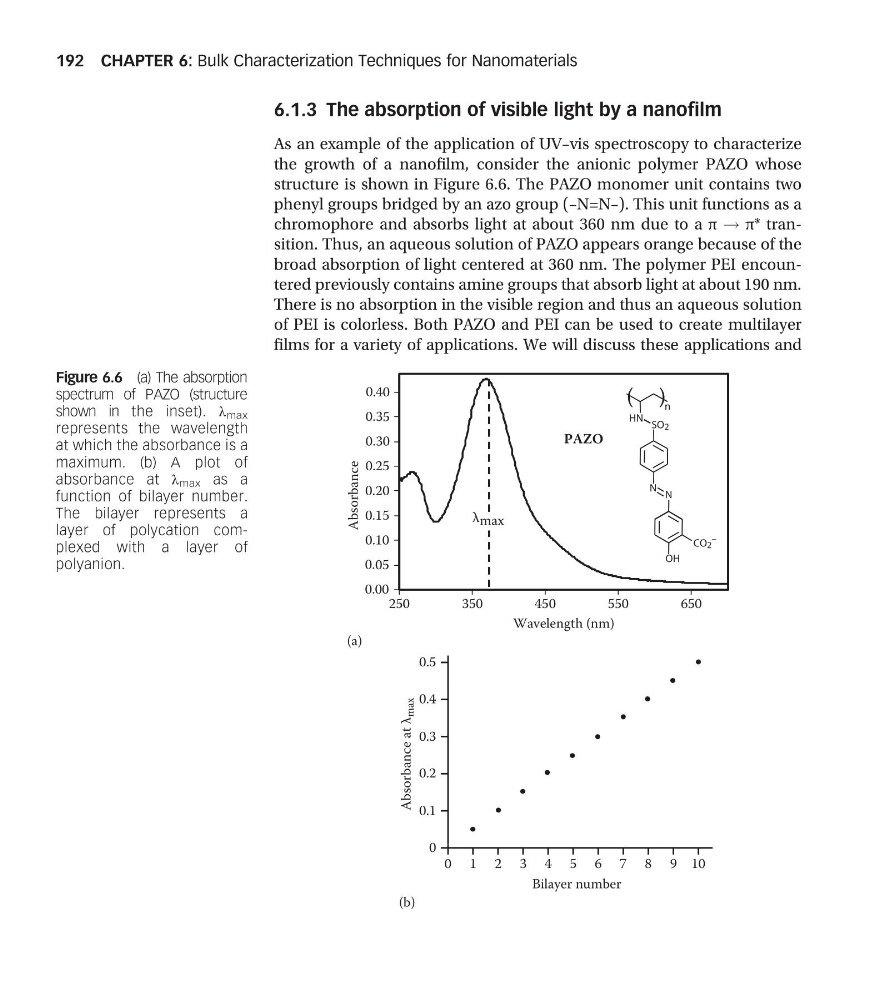
structure is shown in Figure 6.6. The PAZO monomer unit contains two
phenyl groups bridged by an azo group (–N=N–). This unit functions as a
chromophore and absorbs light at about 360 nm due to a π ! π* transition. Thus, an aqueous solution of PAZO appears orange because of the
broad absorption of light centered at 360 nm. The polymer PEI encountered previously contains amine groups that absorb light at about 190 nm.
There is no absorption in the visible region and thus an aqueous solution
of PEI is colorless. Both PAZO and PEI can be used to create multilayer
films for a variety of applications. We will discuss these applications and
0.35
0.40
n
(a)
0.15
0.20
0.25
0.30
N N
SO2
HN
PAZO
0.00
250
350
450
550
650
0.05
0.10
Absorbance
CO2
–
OH
λ max
Wavelength (nm)
0.5
(b)
0.3
0.4
Absorbance at λ
max
0
0.1
0.2
Bilayer number
10
9
8
7
6
5
4
3
2
1
0
Figure 6.6 (a) The absorption
spectrum of PAZO (structure
shown in the inset). λ max
represents the wavelength
at which the absorbance is a
maximum. (b) A plot of
absorbance at λ max as a
function of bilayer number.
The bilayer represents a
layer of polycation complexed with a layer of
polyanion.
CHAPTER 6: Bulk Characterization Techniques for Nanomaterials
192
As an example of the application of UV–vis spectroscopy to characterize
the growth of a nanofilm, consider the anionic polymer PAZO whose
structure is shown in Figure 6.6. The PAZO monomer unit contains two
phenyl groups bridged by an azo group (–N=N–). This unit functions as a
chromophore and absorbs light at about 360 nm due to a π ! π* transition. Thus, an aqueous solution of PAZO appears orange because of the
broad absorption of light centered at 360 nm. The polymer PEI encountered previously contains amine groups that absorb light at about 190 nm.
There is no absorption in the visible region and thus an aqueous solution
of PEI is colorless. Both PAZO and PEI can be used to create multilayer
films for a variety of applications. We will discuss these applications and
0.35
0.40
n
(a)
0.15
0.20
0.25
0.30
N N
SO2
HN
PAZO
0.00
250
350
450
550
650
0.05
0.10
Absorbance
CO2
–
OH
λ max
Wavelength (nm)
0.5
(b)
0.3
0.4
Absorbance at λ
max
0
0.1
0.2
Bilayer number
10
9
8
7
6
5
4
3
2
1
0
Figure 6.6 (a) The absorption
spectrum of PAZO (structure
shown in the inset). λ max
represents the wavelength
at which the absorbance is a
maximum. (b) A plot of
absorbance at λ max as a
function of bilayer number.
The bilayer represents a
layer of polycation complexed with a layer of
polyanion.
CHAPTER 6: Bulk Characterization Techniques for Nanomaterials
192