

Antileishmanial Activity of Lignans, Neolignans …
119
postdisease syndromes or complications connected with VL and CL as post-kalaazar dermal syndrome (PKDL), the mucocutaneous form (MCL), the disseminated
form (DsCL), the diffuse form (DCL), coinfection (AIDS/leishmaniasis), and others
[17]. In general, the widely spread L. donovani (India, Africa regions), L. infantum
(Mediterranean areas) and L. chagasi (Latin America) cause visceral leishmaniasis
(VL). While visceral leishmaniasis is endemic in more than 60 countries, 90% of all
cases are reported from just seven countries: Brazil, Ethiopia, Kenya, Somalia, South
Sudan, Sudan, and India [21]. In the case of L. donovani, the main host reservoir is
the human, and for L. infantum, the domestic dog, but other mammalian reservoirs
also exist [40]. The incubation period of the parasite ranges from 2 weeks up to 8
months, and the beginning of incubation can be sudden, asymptomatic, or variable,
while the disease itself (leishmaniasis) can develop even a year after incubation when
the patient immune system is suppressed. If not treated, VL is fatal and the host is
killed often due to a secondary bacterial infection within 2 years after the parasite
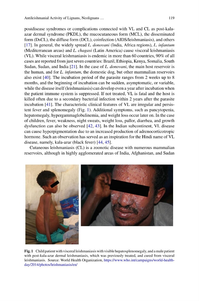
incubation [41]. The characteristic clinical features of VL are irregular and persistent fever and splenomegaly (Fig. 1). Additional symptoms, such as pancytopenia,
hepatomegaly, hypergammaglobulinemia, and weight loss occur later on. In the case
of children, fever, weakness, night sweats, weight loss, pallor, diarrhea, and growth
dysfunction can also be observed [42, 43]. In the Indian subcontinent, VL disease
can cause hyperpigmentation due to an increased production of adrenocorticotropic
hormone. Such an observation has served as an inspiration for the Hindi name of VL
disease, namely, kala-azar (black fever) [44, 45].
Cutaneous leishmaniasis (CL) is a zoonotic disease with numerous mammalian
reservoirs, although in highly agglomerated areas of India, Afghanistan, and Sudan
Fig. 1 Child patient with visceral leishmaniasis with visible hepatosplenomegaly, and a male patient
with post-kala-azar dermal leishmaniasis, which was previously treated, and cured from visceral
leishmaniasis. Source: World Health Organization, https://www.who.int/campaigns/world-healthday/2014/photos/leishmaniasis/en/
119
postdisease syndromes or complications connected with VL and CL as post-kalaazar dermal syndrome (PKDL), the mucocutaneous form (MCL), the disseminated
form (DsCL), the diffuse form (DCL), coinfection (AIDS/leishmaniasis), and others
[17]. In general, the widely spread L. donovani (India, Africa regions), L. infantum
(Mediterranean areas) and L. chagasi (Latin America) cause visceral leishmaniasis
(VL). While visceral leishmaniasis is endemic in more than 60 countries, 90% of all
cases are reported from just seven countries: Brazil, Ethiopia, Kenya, Somalia, South
Sudan, Sudan, and India [21]. In the case of L. donovani, the main host reservoir is
the human, and for L. infantum, the domestic dog, but other mammalian reservoirs
also exist [40]. The incubation period of the parasite ranges from 2 weeks up to 8
months, and the beginning of incubation can be sudden, asymptomatic, or variable,
while the disease itself (leishmaniasis) can develop even a year after incubation when
the patient immune system is suppressed. If not treated, VL is fatal and the host is
killed often due to a secondary bacterial infection within 2 years after the parasite
incubation [41]. The characteristic clinical features of VL are irregular and persistent fever and splenomegaly (Fig. 1). Additional symptoms, such as pancytopenia,
hepatomegaly, hypergammaglobulinemia, and weight loss occur later on. In the case
of children, fever, weakness, night sweats, weight loss, pallor, diarrhea, and growth
dysfunction can also be observed [42, 43]. In the Indian subcontinent, VL disease
can cause hyperpigmentation due to an increased production of adrenocorticotropic
hormone. Such an observation has served as an inspiration for the Hindi name of VL
disease, namely, kala-azar (black fever) [44, 45].
Cutaneous leishmaniasis (CL) is a zoonotic disease with numerous mammalian
reservoirs, although in highly agglomerated areas of India, Afghanistan, and Sudan
Fig. 1 Child patient with visceral leishmaniasis with visible hepatosplenomegaly, and a male patient
with post-kala-azar dermal leishmaniasis, which was previously treated, and cured from visceral
leishmaniasis. Source: World Health Organization, https://www.who.int/campaigns/world-healthday/2014/photos/leishmaniasis/en/