

118
J. Pospíšil et al.
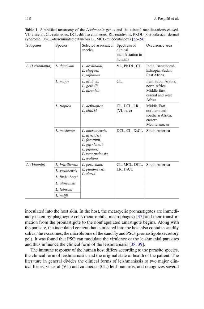
Table 1 Simplified taxonomy of the Leishmania genus and the clinical manifestations caused.
VL-visceral, CL-cutaneous, DCL-diffuse cutaneous, RL-recidivans, PKDL-post-kala-azar dermal
syndrome, DsCL-disseminated cutaneous L., MCL-mucocutaneous [22–24]
Subgenus
Species
Selected associated
species
Spectrum of
clinical
manifestation in
humans
Occurrence area
L. (Leishmania) L. donovani
L. archibaldi,
L. chagasi,
L. infantum
VL, PKDL, CL India, Bangladesh,
Ethiopia, Sudan,
East Africa
L. major
L. arabica,
L. gerbilli,
L. turanica
CL
Iran, Saudi Arabia,
north Africa,
Middle East,
central and west
Africa
L. tropica
L. aethiopica,
L. killicki
CL, DCL, LR,
(VL-rare)
Middle East,
northern and
southern Africa,
eastern
Mediterranean
L. mexicana
L. amazonensis,
L. aristidesi,
L. forattinii,
L. garnhamii,
L. pifanoi,
L. venezuelensis,
L. waltoni
DCL, CL, DsCL South America
L. (Viannia)
L. braziliensis L. peruviana,
L. panamensis,
L. shawi
CL, MCL, DCL,
LR, DsCl,
South America
L. guyanensis
L. lindenbergi
L. utingensis
L. lainsoni
L. naiffi
inoculated into the host skin. In the host, the metacyclic promastigotes are immediately taken by phagocytic cells (neutrophils, macrophages) [37] and their transformation from the promastigote to the nonflagellated amastigote begins. Along with
the parasite, the inoculated content that is injected into the host also contains sandfly
saliva, the exosomes, the microbiome of the sand fly and PSG (promastigote secretory
gel). It was found that PSG can modulate the virulence of the leishmanial parasites
and thus influence the clinical form of the leishmaniasis [38, 39].
The immune response of the human host differs according to the parasite species,
the clinical form of leishmaniasis, and the original state of health of the patient. The
literature in general divides the clinical forms of leishmaniasis to two major clinical forms, visceral (VL) and cutaneous (CL) leishmaniasis, and recognizes several
J. Pospíšil et al.
Table 1 Simplified taxonomy of the Leishmania genus and the clinical manifestations caused.
VL-visceral, CL-cutaneous, DCL-diffuse cutaneous, RL-recidivans, PKDL-post-kala-azar dermal
syndrome, DsCL-disseminated cutaneous L., MCL-mucocutaneous [22–24]
Subgenus
Species
Selected associated
species
Spectrum of
clinical
manifestation in
humans
Occurrence area
L. (Leishmania) L. donovani
L. archibaldi,
L. chagasi,
L. infantum
VL, PKDL, CL India, Bangladesh,
Ethiopia, Sudan,
East Africa
L. major
L. arabica,
L. gerbilli,
L. turanica
CL
Iran, Saudi Arabia,
north Africa,
Middle East,
central and west
Africa
L. tropica
L. aethiopica,
L. killicki
CL, DCL, LR,
(VL-rare)
Middle East,
northern and
southern Africa,
eastern
Mediterranean
L. mexicana
L. amazonensis,
L. aristidesi,
L. forattinii,
L. garnhamii,
L. pifanoi,
L. venezuelensis,
L. waltoni
DCL, CL, DsCL South America
L. (Viannia)
L. braziliensis L. peruviana,
L. panamensis,
L. shawi
CL, MCL, DCL,
LR, DsCl,
South America
L. guyanensis
L. lindenbergi
L. utingensis
L. lainsoni
L. naiffi
inoculated into the host skin. In the host, the metacyclic promastigotes are immediately taken by phagocytic cells (neutrophils, macrophages) [37] and their transformation from the promastigote to the nonflagellated amastigote begins. Along with
the parasite, the inoculated content that is injected into the host also contains sandfly
saliva, the exosomes, the microbiome of the sand fly and PSG (promastigote secretory
gel). It was found that PSG can modulate the virulence of the leishmanial parasites
and thus influence the clinical form of the leishmaniasis [38, 39].
The immune response of the human host differs according to the parasite species,
the clinical form of leishmaniasis, and the original state of health of the patient. The
literature in general divides the clinical forms of leishmaniasis to two major clinical forms, visceral (VL) and cutaneous (CL) leishmaniasis, and recognizes several