

12.3 Electron Microscopy 301
In particular, the localized chemical analysis using X-rays or EELS allow the identification of lattice elements or even single atoms.
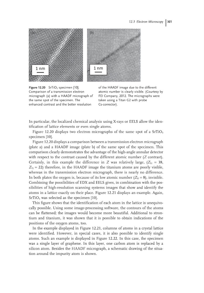
Figure 12.20 displays two electron micrographs of the same spot of a SrTiO 3
specimen [10].
Figure 12.20 displays a comparison between a transmission electron micrograph
(plate a) and a HAADF image (plate b) of the same spot of the specimen. This
comparison clearly demonstrates the advantage of the high-angle annular detector
with respect to the contrast caused by the different atomic number (Z contrast).
Certainly, in this example the difference in Z was relatively large, (Z Sr = 38,
Z Ti = 22) therefore, in the HAADF image the titanium atoms are poorly visible,
whereas in the transmission electron micrograph, there is nearly no difference.
In both plates the oxygen is, because of its low atomic number (Z O = 8), invisible.
Combining the possibilities of EDX and EELS gives, in combination with the possibilities of high-resolution scanning systems images that show and identify the
atoms in a lattice exactly on their place. Figure 12.21 displays an example. Again,
SrTiO 3 was selected as the specimen [10].
This figure shows that the identification of each atom in the lattice is unequivocally possible. Using some image-processing software, the contours of the atoms
can be flattened; the images would become more beautiful. Additional to strontium and titanium, it was shown that it is possible to obtain indications of the
positions of the oxygen atoms, too.
In the example displayed in Figure 12.21, columns of atoms in a crystal lattice
were identified. However, in special cases, it is also possible to identify single
atoms. Such an example is displayed in Figure 12.22. In this case, the specimen
was a single layer of graphene. In this layer, one carbon atom is replaced by a
silicon atom. Besides the HAADF micrograph, a schematic drawing of the situation around the impurity atom is shown.
Figure 12.20 SrTiO 3 specimen [10].
Comparison of a transmission electron
micrograph (a) with a HAADF micrograph of
the same spot of the specimen. The
enhanced contrast and the better resolution
of the HAADF image due to the different
atomic number is clearly visible. (Courtesy by
FEI Company, 2012. The micrographs were
taken using a Titan G2 with probe
Cs-corrector).
1 nm
1 nm
(a)
(b)
In particular, the localized chemical analysis using X-rays or EELS allow the identification of lattice elements or even single atoms.
Figure 12.20 displays two electron micrographs of the same spot of a SrTiO 3
specimen [10].
Figure 12.20 displays a comparison between a transmission electron micrograph
(plate a) and a HAADF image (plate b) of the same spot of the specimen. This
comparison clearly demonstrates the advantage of the high-angle annular detector
with respect to the contrast caused by the different atomic number (Z contrast).
Certainly, in this example the difference in Z was relatively large, (Z Sr = 38,
Z Ti = 22) therefore, in the HAADF image the titanium atoms are poorly visible,
whereas in the transmission electron micrograph, there is nearly no difference.
In both plates the oxygen is, because of its low atomic number (Z O = 8), invisible.
Combining the possibilities of EDX and EELS gives, in combination with the possibilities of high-resolution scanning systems images that show and identify the
atoms in a lattice exactly on their place. Figure 12.21 displays an example. Again,
SrTiO 3 was selected as the specimen [10].
This figure shows that the identification of each atom in the lattice is unequivocally possible. Using some image-processing software, the contours of the atoms
can be flattened; the images would become more beautiful. Additional to strontium and titanium, it was shown that it is possible to obtain indications of the
positions of the oxygen atoms, too.
In the example displayed in Figure 12.21, columns of atoms in a crystal lattice
were identified. However, in special cases, it is also possible to identify single
atoms. Such an example is displayed in Figure 12.22. In this case, the specimen
was a single layer of graphene. In this layer, one carbon atom is replaced by a
silicon atom. Besides the HAADF micrograph, a schematic drawing of the situation around the impurity atom is shown.
Figure 12.20 SrTiO 3 specimen [10].
Comparison of a transmission electron
micrograph (a) with a HAADF micrograph of
the same spot of the specimen. The
enhanced contrast and the better resolution
of the HAADF image due to the different
atomic number is clearly visible. (Courtesy by
FEI Company, 2012. The micrographs were
taken using a Titan G2 with probe
Cs-corrector).
1 nm
1 nm
(a)
(b)