

300 12 Characterization of Nanomaterials
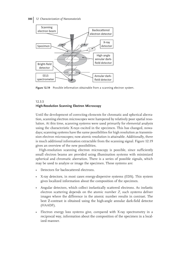
Figure 12.19 Possible information obtainable from a scanning electron system.
Scanning
electron beam
BackscaƩered
electron detector
X-ray
detector
High-angle
annular darkfield detector
Annular darkfield detector
Bright-field
detector
Specimen
EELS
spectrometer
12.3.5
High-Resolution Scanning Electron Microscopy
Until the development of correcting elements for chromatic and spherical aberration, scanning electron microscopes were hampered by relatively poor spatial resolution. At this time, scanning systems were used primarily for elemental analysis
using the characteristic X-rays excited in the specimen. This has changed, nowadays; scanning systems have the same possibilities for high resolution as transmission electron microscopes; now atomic resolution is attainable. Additionally, there
is much additional information extractable from the scanning signal. Figure 12.19
gives an overview of the new possibilities.
High-resolution scanning electron microscopy is possible, since sufficiently
small electron beams are provided using illumination systems with minimized
spherical and chromatic aberration. There is a series of possible signals, which
may be used to analyze or image the specimen. These systems are:
• Detectors for backscattered electrons.
• X-ray detectors, in most cases energy-dispersive systems (EDX). This system
gives localized information about the composition of the specimen.
• Angular detectors, which collect inelastically scattered electrons. As inelastic
electron scattering depends on the atomic number Z, such systems deliver
images where the difference in the atomic number results in contrast. The
best Z-contrast is obtained using the high-angle annular dark-field detector
(HAADF).
• Electron energy loss systems give, compared with X-ray spectrometry in a
reciprocal way, information about the composition of the specimen in a localized manner.
Figure 12.19 Possible information obtainable from a scanning electron system.
Scanning
electron beam
BackscaƩered
electron detector
X-ray
detector
High-angle
annular darkfield detector
Annular darkfield detector
Bright-field
detector
Specimen
EELS
spectrometer
12.3.5
High-Resolution Scanning Electron Microscopy
Until the development of correcting elements for chromatic and spherical aberration, scanning electron microscopes were hampered by relatively poor spatial resolution. At this time, scanning systems were used primarily for elemental analysis
using the characteristic X-rays excited in the specimen. This has changed, nowadays; scanning systems have the same possibilities for high resolution as transmission electron microscopes; now atomic resolution is attainable. Additionally, there
is much additional information extractable from the scanning signal. Figure 12.19
gives an overview of the new possibilities.
High-resolution scanning electron microscopy is possible, since sufficiently
small electron beams are provided using illumination systems with minimized
spherical and chromatic aberration. There is a series of possible signals, which
may be used to analyze or image the specimen. These systems are:
• Detectors for backscattered electrons.
• X-ray detectors, in most cases energy-dispersive systems (EDX). This system
gives localized information about the composition of the specimen.
• Angular detectors, which collect inelastically scattered electrons. As inelastic
electron scattering depends on the atomic number Z, such systems deliver
images where the difference in the atomic number results in contrast. The
best Z-contrast is obtained using the high-angle annular dark-field detector
(HAADF).
• Electron energy loss systems give, compared with X-ray spectrometry in a
reciprocal way, information about the composition of the specimen in a localized manner.