

12.3 Electron Microscopy 299
Figure 12.17 High-resolution electron
micrograph of a graphene sheet. This
micrograph was taken with an electron
energy of 80 keV [9]. One sees the elementary
structure of graphene, the hexagons, which
are highlighted in one example in the white
circle. The distance between two of these
points is 0.14 nm. (Courtesy by FEI Company,
2012).
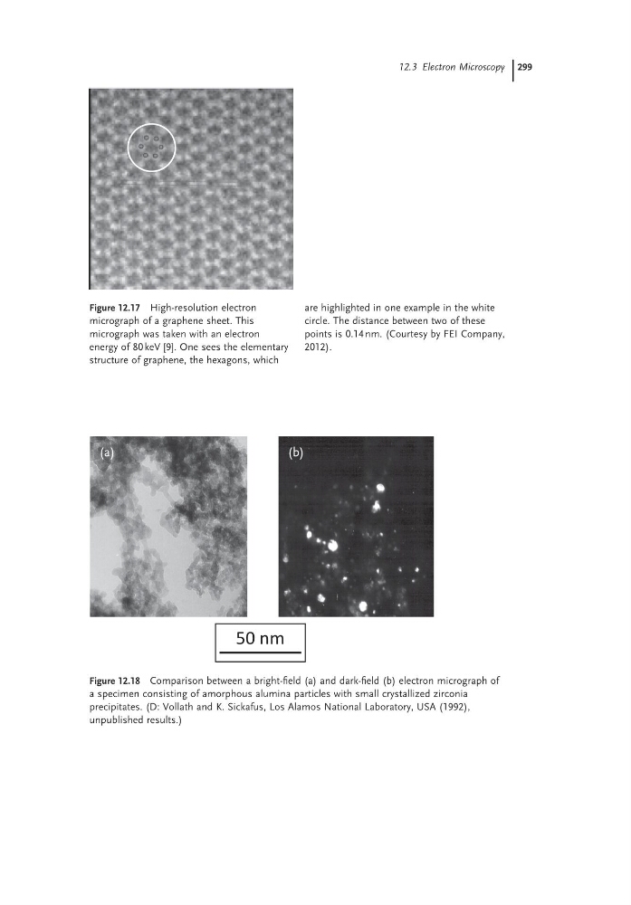
Figure 12.18 Comparison between a bright-field (a) and dark-field (b) electron micrograph of
a specimen consisting of amorphous alumina particles with small crystallized zirconia
precipitates. (D: Vollath and K. Sickafus, Los Alamos National Laboratory, USA (1992),
unpublished results.)
50 nm
(a)
(b)
Figure 12.17 High-resolution electron
micrograph of a graphene sheet. This
micrograph was taken with an electron
energy of 80 keV [9]. One sees the elementary
structure of graphene, the hexagons, which
are highlighted in one example in the white
circle. The distance between two of these
points is 0.14 nm. (Courtesy by FEI Company,
2012).
Figure 12.18 Comparison between a bright-field (a) and dark-field (b) electron micrograph of
a specimen consisting of amorphous alumina particles with small crystallized zirconia
precipitates. (D: Vollath and K. Sickafus, Los Alamos National Laboratory, USA (1992),
unpublished results.)
50 nm
(a)
(b)