

9.6 Luminescent Nanocomposites 205
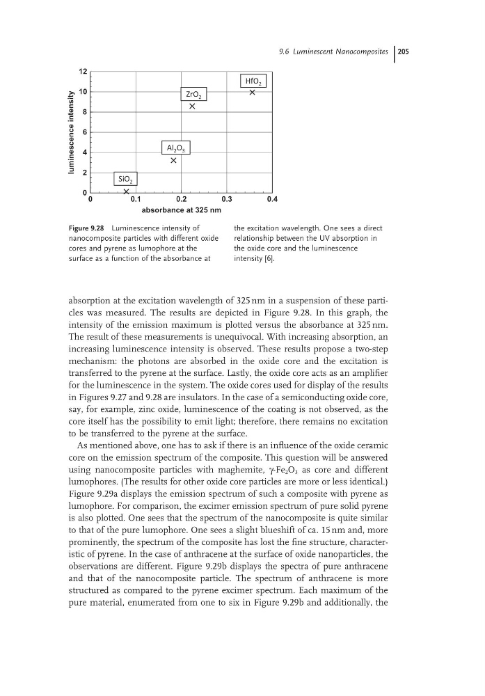
absorption at the excitation wavelength of 325 nm in a suspension of these particles was measured. The results are depicted in Figure 9.28. In this graph, the
intensity of the emission maximum is plotted versus the absorbance at 325 nm.
The result of these measurements is unequivocal. With increasing absorption, an
increasing luminescence intensity is observed. These results propose a two-step
mechanism: the photons are absorbed in the oxide core and the excitation is
transferred to the pyrene at the surface. Lastly, the oxide core acts as an amplifier
for the luminescence in the system. The oxide cores used for display of the results
in Figures 9.27 and 9.28 are insulators. In the case of a semiconducting oxide core,
say, for example, zinc oxide, luminescence of the coating is not observed, as the
core itself has the possibility to emit light; therefore, there remains no excitation
to be transferred to the pyrene at the surface.
As mentioned above, one has to ask if there is an influence of the oxide ceramic
core on the emission spectrum of the composite. This question will be answered
using nanocomposite particles with maghemite, γ-Fe 2 O 3 as core and different
lumophores. (The results for other oxide core particles are more or less identical.)
Figure 9.29a displays the emission spectrum of such a composite with pyrene as
lumophore. For comparison, the excimer emission spectrum of pure solid pyrene
is also plotted. One sees that the spectrum of the nanocomposite is quite similar
to that of the pure lumophore. One sees a slight blueshift of ca. 15 nm and, more
prominently, the spectrum of the composite has lost the fine structure, characteristic of pyrene. In the case of anthracene at the surface of oxide nanoparticles, the
observations are different. Figure 9.29b displays the spectra of pure anthracene
and that of the nanocomposite particle. The spectrum of anthracene is more
structured as compared to the pyrene excimer spectrum. Each maximum of the
pure material, enumerated from one to six in Figure 9.29b and additionally, the
Figure 9.28 Luminescence intensity of
nanocomposite particles with different oxide
cores and pyrene as lumophore at the
surface as a function of the absorbance at
the excitation wavelength. One sees a direct
relationship between the UV absorption in
the oxide core and the luminescence
intensity [6].
0
0.1
0.2
0.3
0.4
absorbance at 325 nm
0
2
4
6
8
10
12
luminescence
intensity
HfO 2
ZrO 2
Al 2 O 3
SiO 2
absorption at the excitation wavelength of 325 nm in a suspension of these particles was measured. The results are depicted in Figure 9.28. In this graph, the
intensity of the emission maximum is plotted versus the absorbance at 325 nm.
The result of these measurements is unequivocal. With increasing absorption, an
increasing luminescence intensity is observed. These results propose a two-step
mechanism: the photons are absorbed in the oxide core and the excitation is
transferred to the pyrene at the surface. Lastly, the oxide core acts as an amplifier
for the luminescence in the system. The oxide cores used for display of the results
in Figures 9.27 and 9.28 are insulators. In the case of a semiconducting oxide core,
say, for example, zinc oxide, luminescence of the coating is not observed, as the
core itself has the possibility to emit light; therefore, there remains no excitation
to be transferred to the pyrene at the surface.
As mentioned above, one has to ask if there is an influence of the oxide ceramic
core on the emission spectrum of the composite. This question will be answered
using nanocomposite particles with maghemite, γ-Fe 2 O 3 as core and different
lumophores. (The results for other oxide core particles are more or less identical.)
Figure 9.29a displays the emission spectrum of such a composite with pyrene as
lumophore. For comparison, the excimer emission spectrum of pure solid pyrene
is also plotted. One sees that the spectrum of the nanocomposite is quite similar
to that of the pure lumophore. One sees a slight blueshift of ca. 15 nm and, more
prominently, the spectrum of the composite has lost the fine structure, characteristic of pyrene. In the case of anthracene at the surface of oxide nanoparticles, the
observations are different. Figure 9.29b displays the spectra of pure anthracene
and that of the nanocomposite particle. The spectrum of anthracene is more
structured as compared to the pyrene excimer spectrum. Each maximum of the
pure material, enumerated from one to six in Figure 9.29b and additionally, the
Figure 9.28 Luminescence intensity of
nanocomposite particles with different oxide
cores and pyrene as lumophore at the
surface as a function of the absorbance at
the excitation wavelength. One sees a direct
relationship between the UV absorption in
the oxide core and the luminescence
intensity [6].
0
0.1
0.2
0.3
0.4
absorbance at 325 nm
0
2
4
6
8
10
12
luminescence
intensity
HfO 2
ZrO 2
Al 2 O 3
SiO 2