

9. Once the scaffold printing is complete, switch off voltage followed by gas pressure and temperature.
10. Turn off machine, peel off scaffold, and store safely. Clean any
remaining sample on the syringe tip and collector with ethanol/acetone and paper towel.
3.2 Preparing the
Scaffolds for Cell
Culture
1. Use a biopsy punch or laser cutter to cut the scaffold to the
desired size.
2. Sterilize the scaffolds by soaking them in 80% ethanol for
30 min, followed by UV irradiation for 20 min on each side.
Table 2
(continued)
o5
g1 x0 y60.00 f650
g02 x0.200 y0 r–0.400 f170
g1 x0 y–60.00 f650
g03 x0.200 y0 r–0.400 f170
m99
Subprogram “5,” the stage moves 60 units in the positive y direction at a feed rate of 650 mm/min. The
stage then does a loop with a radius of 0.4 mm, moving clockwise and leaving an opening of 0.2 mm.
The feed rate here is f170 mm/min. The stage then moves 60 mm in the negative y direction and does
a counterclockwise loop. m99 ends the subprogram
o6
g1 x0 y60.00 f650
m99
Subprogram “6,” the stage moves 60 mm in the positive y direction at a feed rate of 650 mm/min
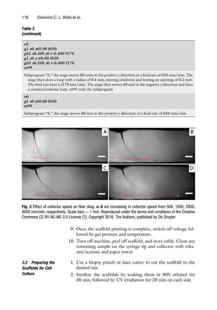
Fig. 3 Effect of collector speed on fiber drag. a–d are increasing in collector speed from 500, 1000, 2000,
8000 mm/min, respectively. Scale bars ¼ 1 mm. Reproduced under the terms and conditions of the Creative
Commons CC BY-NC-ND 3.0 License [7]. Copyright 2016, The Authors, published by De Gruyter
118
Eleonore C. L. Bolle et al.
10. Turn off machine, peel off scaffold, and store safely. Clean any
remaining sample on the syringe tip and collector with ethanol/acetone and paper towel.
3.2 Preparing the
Scaffolds for Cell
Culture
1. Use a biopsy punch or laser cutter to cut the scaffold to the
desired size.
2. Sterilize the scaffolds by soaking them in 80% ethanol for
30 min, followed by UV irradiation for 20 min on each side.
Table 2
(continued)
o5
g1 x0 y60.00 f650
g02 x0.200 y0 r–0.400 f170
g1 x0 y–60.00 f650
g03 x0.200 y0 r–0.400 f170
m99
Subprogram “5,” the stage moves 60 units in the positive y direction at a feed rate of 650 mm/min. The
stage then does a loop with a radius of 0.4 mm, moving clockwise and leaving an opening of 0.2 mm.
The feed rate here is f170 mm/min. The stage then moves 60 mm in the negative y direction and does
a counterclockwise loop. m99 ends the subprogram
o6
g1 x0 y60.00 f650
m99
Subprogram “6,” the stage moves 60 mm in the positive y direction at a feed rate of 650 mm/min
Fig. 3 Effect of collector speed on fiber drag. a–d are increasing in collector speed from 500, 1000, 2000,
8000 mm/min, respectively. Scale bars ¼ 1 mm. Reproduced under the terms and conditions of the Creative
Commons CC BY-NC-ND 3.0 License [7]. Copyright 2016, The Authors, published by De Gruyter
118
Eleonore C. L. Bolle et al.