

3. Centrifuge the scaffolds in fibroblasts growth medium at
400 Â g for 10 min to fully immerse the scaffolds in medium
and to minimize the air trapped inside the scaffold.
4. Place the scaffolds in fibroblasts growth medium and keep
them in the incubator overnight (see Note 4).
3.3 Seeding the
Scaffolds with Cells
1. Once the cells reach 80% confluence, they can be seeded onto
the scaffolds.
2. Remove the culture medium from the flask and rinse cells with
5 mL of sterile PBS.
3. Add 1–2 mL of trypsin to the flask and incubate for 2 min to
detach the cells from the flask.
4. Once the cells begin to retract, tap the sides of the flask to fully
detach the cells.
5. Add 10 mL of fibroblasts medium to inhibit the trypsin, wash
down the flask with the medium, and transfer the cell suspension into a falcon tube.
6. Spin down the cell suspension at 200 Â g for 5 min, and
resuspend the cell pellet in 10 mL of fibroblasts medium.
7. Perform a cell count to determine the amount of viable cells.
Dilute 100 μL of cell suspension in 100 μL of trypan blue, and
add 10 μL of the dilution into the hemocytometer.
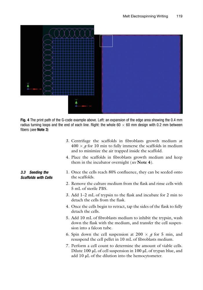
Fig. 4 The print path of the G-code example above. Left: an expansion of the edge area showing the 0.4 mm
radius turning loops and the end of each line. Right: the whole 60 Â 60 mm design with 0.2 mm between
fibers (see Note 3)
Melt Electrospinning Writing
119
400 Â g for 10 min to fully immerse the scaffolds in medium
and to minimize the air trapped inside the scaffold.
4. Place the scaffolds in fibroblasts growth medium and keep
them in the incubator overnight (see Note 4).
3.3 Seeding the
Scaffolds with Cells
1. Once the cells reach 80% confluence, they can be seeded onto
the scaffolds.
2. Remove the culture medium from the flask and rinse cells with
5 mL of sterile PBS.
3. Add 1–2 mL of trypsin to the flask and incubate for 2 min to
detach the cells from the flask.
4. Once the cells begin to retract, tap the sides of the flask to fully
detach the cells.
5. Add 10 mL of fibroblasts medium to inhibit the trypsin, wash
down the flask with the medium, and transfer the cell suspension into a falcon tube.
6. Spin down the cell suspension at 200 Â g for 5 min, and
resuspend the cell pellet in 10 mL of fibroblasts medium.
7. Perform a cell count to determine the amount of viable cells.
Dilute 100 μL of cell suspension in 100 μL of trypan blue, and
add 10 μL of the dilution into the hemocytometer.
Fig. 4 The print path of the G-code example above. Left: an expansion of the edge area showing the 0.4 mm
radius turning loops and the end of each line. Right: the whole 60 Â 60 mm design with 0.2 mm between
fibers (see Note 3)
Melt Electrospinning Writing
119