

716
Z. Voitko et al.
detected [17]. The main peak of pure collagen is 2856 cm
−1 , which corresponds to
the asymmetric and symmetric CH stretching modes [18].
Raman spectroscopy results are in a good agreement with X-ray diffraction and
IR spectroscopy results.
2.7 Microscopy
Scanning electron microscopy studies were performed for samples of HA, HA–
collagen, and HA–collagen–alginate composite. The latter was obtained in the form
of balls to increase overall biocompatibility, facilitate storage, and use.
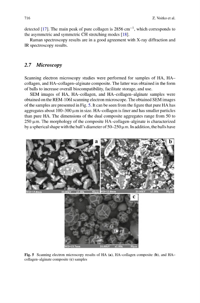
SEM images of HA, HA–collagen, and HA–collagen–alginate samples were
obtained on the REM-106I scanning electron microscope. The obtained SEM images
of the samples are presented in Fig. 5. It can be seen from the figure that pure HA has
aggregates about 100–300 µm in size. HA–collagen is finer and has smaller particles
than pure HA. The dimensions of the dual composite aggregates range from 50 to
250 µm. The morphology of the composite HA–collagen–alginate is characterized
by a spherical shape with the ball’s diameter of 50–250 µm. In addition, the balls have
Fig. 5 Scanning electron microscopy results of HA (a), HA–collagen composite (b), and HA–
collagen–alginate composite (c) samples