

268
Biologically Inspired Robotics
the input to the cuneate neuron via a synapse (Figure 14.1a), displays spike
only on stimulation. Therefore, the time before the stimulation is devoid of
spikes, even though the spike response to the stimulation is quite intense
(Figure 14.1b, top). In contrast, the cuneate neuron has a spontaneous activity,
and hence the spike activity before the stimulation, and in addition it seems
to have a more intense spike response to stimulation (note the steep increase
in spike activity immediately after the onset of the stimulation; Figure 14.1b,
bottom).
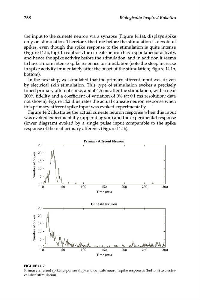
In the next step, we simulated that the primary afferent input was driven
by electrical skin stimulation. This type of stimulation evokes a precisely
timed primary afferent spike, about 4.5 ms after the stimulation, with a near
100% fidelity and a coefficient of variation of 0% (at 0.1 ms resolution; data
not shown). Figure 14.2 illustrates the actual cuneate neuron response when
this primary afferent spike input was evoked experimentally.
Figure 14.2 illustrates the actual cuneate neuron response when this input
was evoked experimentally (upper diagram) and the experimental response
(lower diagram) evoked by a single pulse input comparable to the spike
response of the real primary afferents (Figure 14.1b).
Primary Afferent Neuron
0
5 0
100
150
200
250
300
Time (ms)
0
5 0
0
5
10
15
20
25
100
Time (ms)
Number of Spikes
150
200
300
250
Cuneate Neuron
0
5
10
15
20
25
Number of Spikes
FIGURE 14.2
Primary afferent spike responses (top) and cuneate neuron spike responses (bottom) to electrical skin stimulation.
Biologically Inspired Robotics
the input to the cuneate neuron via a synapse (Figure 14.1a), displays spike
only on stimulation. Therefore, the time before the stimulation is devoid of
spikes, even though the spike response to the stimulation is quite intense
(Figure 14.1b, top). In contrast, the cuneate neuron has a spontaneous activity,
and hence the spike activity before the stimulation, and in addition it seems
to have a more intense spike response to stimulation (note the steep increase
in spike activity immediately after the onset of the stimulation; Figure 14.1b,
bottom).
In the next step, we simulated that the primary afferent input was driven
by electrical skin stimulation. This type of stimulation evokes a precisely
timed primary afferent spike, about 4.5 ms after the stimulation, with a near
100% fidelity and a coefficient of variation of 0% (at 0.1 ms resolution; data
not shown). Figure 14.2 illustrates the actual cuneate neuron response when
this primary afferent spike input was evoked experimentally.
Figure 14.2 illustrates the actual cuneate neuron response when this input
was evoked experimentally (upper diagram) and the experimental response
(lower diagram) evoked by a single pulse input comparable to the spike
response of the real primary afferents (Figure 14.1b).
Primary Afferent Neuron
0
5 0
100
150
200
250
300
Time (ms)
0
5 0
0
5
10
15
20
25
100
Time (ms)
Number of Spikes
150
200
300
250
Cuneate Neuron
0
5
10
15
20
25
Number of Spikes
FIGURE 14.2
Primary afferent spike responses (top) and cuneate neuron spike responses (bottom) to electrical skin stimulation.