

267
Mathematical Modeling of Brain Circuitry
Distal
index digit
skin
Skin sensor
receptive field
Primary afferent
Cuneate neuron
Synapse
(a)
–100
0
100
200
300
Time (ms)
Time (ms)
Primary afferent
spike responses
0
4
8
12
16
20
Cuneate neuron
spike responses
–100
0
0
4
8
12
16
20
100
200
300
(b)
FIGURE 14.1
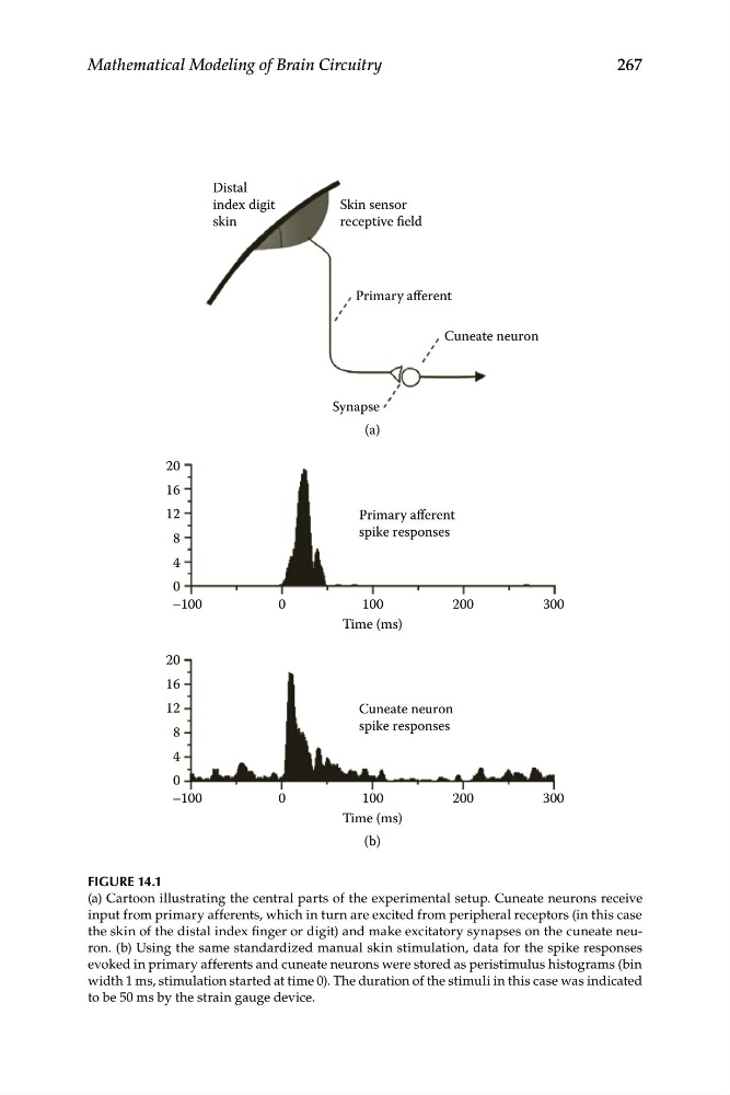
(a) Cartoon illustrating the central parts of the experimental setup. Cuneate neurons receive
input from primary afferents, which in turn are excited from peripheral receptors (in this case
the skin of the distal index finger or digit) and make excitatory synapses on the cuneate neuron. (b) Using the same standardized manual skin stimulation, data for the spike responses
evoked in primary afferents and cuneate neurons were stored as peristimulus histograms (bin
width 1 ms, stimulation started at time 0). The duration of the stimuli in this case was indicated
to be 50 ms by the strain gauge device.
Mathematical Modeling of Brain Circuitry
Distal
index digit
skin
Skin sensor
receptive field
Primary afferent
Cuneate neuron
Synapse
(a)
–100
0
100
200
300
Time (ms)
Time (ms)
Primary afferent
spike responses
0
4
8
12
16
20
Cuneate neuron
spike responses
–100
0
0
4
8
12
16
20
100
200
300
(b)
FIGURE 14.1
(a) Cartoon illustrating the central parts of the experimental setup. Cuneate neurons receive
input from primary afferents, which in turn are excited from peripheral receptors (in this case
the skin of the distal index finger or digit) and make excitatory synapses on the cuneate neuron. (b) Using the same standardized manual skin stimulation, data for the spike responses
evoked in primary afferents and cuneate neurons were stored as peristimulus histograms (bin
width 1 ms, stimulation started at time 0). The duration of the stimuli in this case was indicated
to be 50 ms by the strain gauge device.