

197
Measurement of Brain Activity Using Optical and Electrical Methods
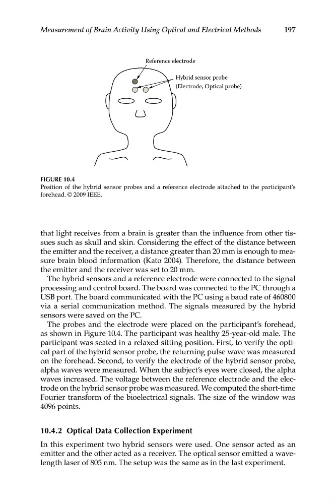
Reference electrode
Hybrid sensor probe
(Electrode, Optical probe)
FIGURE 10.4
Position of the hybrid sensor probes and a reference electrode attached to the participant’s
forehead. © 2009 IEEE.
that light receives from a brain is greater than the influence from other tissues such as skull and skin. Considering the effect of the distance between
the emitter and the receiver, a distance greater than 20 mm is enough to measure brain blood information (Kato 2004). Therefore, the distance between
the emitter and the receiver was set to 20 mm.
The hybrid sensors and a reference electrode were connected to the signal
processing and control board. The board was connected to the PC through a
USB port. The board communicated with the PC using a baud rate of 460800
via a serial communication method. The signals measured by the hybrid
sensors were saved on the PC.
The probes and the electrode were placed on the participant’s forehead,
as shown in Figure 10.4. The participant was healthy 25-year-old male. The
participant was seated in a relaxed sitting position. First, to verify the optical part of the hybrid sensor probe, the returning pulse wave was measured
on the forehead. Second, to verify the electrode of the hybrid sensor probe,
alpha waves were measured. When the subject’s eyes were closed, the alpha
waves increased. The voltage between the reference electrode and the electrode on the hybrid sensor probe was measured. We computed the short-time
Fourier transform of the bioelectrical signals. The size of the window was
4096 points.
10.4.2 Optical Data Collection Experiment
In this experiment two hybrid sensors were used. One sensor acted as an
emitter and the other acted as a receiver. The optical sensor emitted a wavelength laser of 805 nm. The setup was the same as in the last experiment.
Measurement of Brain Activity Using Optical and Electrical Methods
Reference electrode
Hybrid sensor probe
(Electrode, Optical probe)
FIGURE 10.4
Position of the hybrid sensor probes and a reference electrode attached to the participant’s
forehead. © 2009 IEEE.
that light receives from a brain is greater than the influence from other tissues such as skull and skin. Considering the effect of the distance between
the emitter and the receiver, a distance greater than 20 mm is enough to measure brain blood information (Kato 2004). Therefore, the distance between
the emitter and the receiver was set to 20 mm.
The hybrid sensors and a reference electrode were connected to the signal
processing and control board. The board was connected to the PC through a
USB port. The board communicated with the PC using a baud rate of 460800
via a serial communication method. The signals measured by the hybrid
sensors were saved on the PC.
The probes and the electrode were placed on the participant’s forehead,
as shown in Figure 10.4. The participant was healthy 25-year-old male. The
participant was seated in a relaxed sitting position. First, to verify the optical part of the hybrid sensor probe, the returning pulse wave was measured
on the forehead. Second, to verify the electrode of the hybrid sensor probe,
alpha waves were measured. When the subject’s eyes were closed, the alpha
waves increased. The voltage between the reference electrode and the electrode on the hybrid sensor probe was measured. We computed the short-time
Fourier transform of the bioelectrical signals. The size of the window was
4096 points.
10.4.2 Optical Data Collection Experiment
In this experiment two hybrid sensors were used. One sensor acted as an
emitter and the other acted as a receiver. The optical sensor emitted a wavelength laser of 805 nm. The setup was the same as in the last experiment.