

138
Biologically Inspired Robotics
cell through consecutive frames, in some cases the system could not locate
the edges of the fibroblast.
7.3 Experimental Results
7.3.1 Oocyte and Fibroblast Suction
After a number of treatments, explained in the previous section, oocytes
and fibroblasts were dispersed in a random manner in different containers
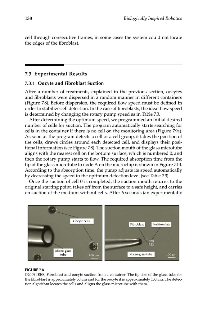
(Figure 7.8). Before dispersion, the required flow speed must be defined in
order to stabilize cell detection. In the case of fibroblasts, the ideal flow speed
is determined by changing the rotary pump speed as in Table 7.3.
After determining the optimum speed, we programmed an initial desired
number of cells for suction. The program automatically starts searching for
cells in the container if there is no cell on the monitoring area (Figure 7.9a).
As soon as the program detects a cell or a cell group, it takes the position of
the cells, draws circles around each detected cell, and displays their positional information (see Figure 7.8). The suction mouth of the glass microtube
aligns with the nearest cell on the bottom surface, which is numbered 0, and
then the rotary pump starts to flow. The required absorption time from the
tip of the glass microtube to node A on the microchip is shown in Figure 7.10.
According to the absorption time, the pump adjusts its speed automatically
by decreasing the speed to the optimum detection level (see Table 7.3).
Once the suction of cell 0 is completed, the suction mouth returns to the
original starting point, takes off from the surface to a safe height, and carries
on suction of the medium without cells. After 6 seconds (an experimentally
Oocyte cells
Micro-glass
tube
100 μm
563
354
77
455
205
35
0
FIGURE 7.8
©2009 IEEE, Fibroblast and oocyte suction from a container. The tip size of the glass tube for
the fibroblast is approximately 50 μm and for the oocyte it is approximately 180 μm. The detection algorithm locates the cells and aligns the glass microtube with them.
Biologically Inspired Robotics
cell through consecutive frames, in some cases the system could not locate
the edges of the fibroblast.
7.3 Experimental Results
7.3.1 Oocyte and Fibroblast Suction
After a number of treatments, explained in the previous section, oocytes
and fibroblasts were dispersed in a random manner in different containers
(Figure 7.8). Before dispersion, the required flow speed must be defined in
order to stabilize cell detection. In the case of fibroblasts, the ideal flow speed
is determined by changing the rotary pump speed as in Table 7.3.
After determining the optimum speed, we programmed an initial desired
number of cells for suction. The program automatically starts searching for
cells in the container if there is no cell on the monitoring area (Figure 7.9a).
As soon as the program detects a cell or a cell group, it takes the position of
the cells, draws circles around each detected cell, and displays their positional information (see Figure 7.8). The suction mouth of the glass microtube
aligns with the nearest cell on the bottom surface, which is numbered 0, and
then the rotary pump starts to flow. The required absorption time from the
tip of the glass microtube to node A on the microchip is shown in Figure 7.10.
According to the absorption time, the pump adjusts its speed automatically
by decreasing the speed to the optimum detection level (see Table 7.3).
Once the suction of cell 0 is completed, the suction mouth returns to the
original starting point, takes off from the surface to a safe height, and carries
on suction of the medium without cells. After 6 seconds (an experimentally
Oocyte cells
Micro-glass
tube
100 μm
563
354
77
455
205
35
0
FIGURE 7.8
©2009 IEEE, Fibroblast and oocyte suction from a container. The tip size of the glass tube for
the fibroblast is approximately 50 μm and for the oocyte it is approximately 180 μm. The detection algorithm locates the cells and aligns the glass microtube with them.