

137
11 Pixels
Pixel Profile
190
170
27 pixels
150
130
Values
110
Pixel
90
70
Pixels
Intensity
50
1 4 7 10 13 16
31 34 37 40 43 46 49 52 55
19 22 25 28
Pixel Number
Automatic Single-Cell Transfer Module
It is important to note that the relative size of each pixel in a digital image
is very important. To determine accurate diameter, the ratio between a
single-cell image reconstructed on an image sensor and corresponding
total pixel number must be calculated. The pixel ratio for each cell was
calibrated by Olympus Ronchi Ruling glass (Olympus, Toyko, Japan) (100
lp/mm). A donor cell of ~15 μm corresponds to about 30 pixels. In addition,
we consider the boundary of any moving region as an edge of the object,
because cells always come into the observation area one by one. Once the
background is fixed, any moving object is filtered according to its size and
dimensions and labeled as a donor or oocyte. Thirty pixels are considered
as a minimum detection size and an object below 30 pixels is omitted. We
essentially applied the same idea to the two camera systems, but cells are
only floating in the container viewed by the microscopic camera. In the
case of the compact vision system, however, the program was plagued by
some environmental problems such as bubbles and other unwanted objects,
which appear similar to the fibroblast or the oocyte.
Becaus the oocytes are approximately ten times larger than the fibroblasts,
requiring a deep channel depth (150 μm), the fibroblasts can easily go out of
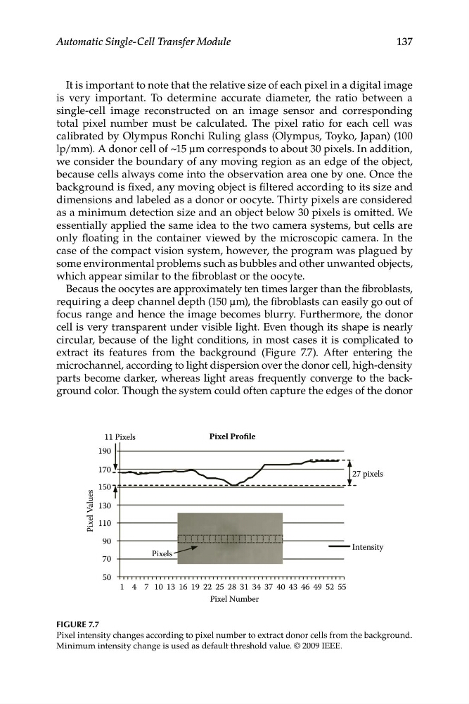
focus range and hence the image becomes blurry. Furthermore, the donor
cell is very transparent under visible light. Even though its shape is nearly
circular, because of the light conditions, in most cases it is complicated to
extract its features from the background (Figure 7.7). After entering the
microchannel, according to light dispersion over the donor cell, high-density
parts become darker, whereas light areas frequently converge to the background color. Though the system could often capture the edges of the donor
FIGURE 7.7
Pixel intensity changes according to pixel number to extract donor cells from the background.
Minimum intensity change is used as default threshold value. © 2009 IEEE.
11 Pixels
Pixel Profile
190
170
27 pixels
150
130
Values
110
Pixel
90
70
Pixels
Intensity
50
1 4 7 10 13 16
31 34 37 40 43 46 49 52 55
19 22 25 28
Pixel Number
Automatic Single-Cell Transfer Module
It is important to note that the relative size of each pixel in a digital image
is very important. To determine accurate diameter, the ratio between a
single-cell image reconstructed on an image sensor and corresponding
total pixel number must be calculated. The pixel ratio for each cell was
calibrated by Olympus Ronchi Ruling glass (Olympus, Toyko, Japan) (100
lp/mm). A donor cell of ~15 μm corresponds to about 30 pixels. In addition,
we consider the boundary of any moving region as an edge of the object,
because cells always come into the observation area one by one. Once the
background is fixed, any moving object is filtered according to its size and
dimensions and labeled as a donor or oocyte. Thirty pixels are considered
as a minimum detection size and an object below 30 pixels is omitted. We
essentially applied the same idea to the two camera systems, but cells are
only floating in the container viewed by the microscopic camera. In the
case of the compact vision system, however, the program was plagued by
some environmental problems such as bubbles and other unwanted objects,
which appear similar to the fibroblast or the oocyte.
Becaus the oocytes are approximately ten times larger than the fibroblasts,
requiring a deep channel depth (150 μm), the fibroblasts can easily go out of
focus range and hence the image becomes blurry. Furthermore, the donor
cell is very transparent under visible light. Even though its shape is nearly
circular, because of the light conditions, in most cases it is complicated to
extract its features from the background (Figure 7.7). After entering the
microchannel, according to light dispersion over the donor cell, high-density
parts become darker, whereas light areas frequently converge to the background color. Though the system could often capture the edges of the donor
FIGURE 7.7
Pixel intensity changes according to pixel number to extract donor cells from the background.
Minimum intensity change is used as default threshold value. © 2009 IEEE.