

84
4 Reactive Thin Polymer Films as Platforms for the Immobilization …
growth capabilities. For instance, listeria can grow, albeit slowly, at temperatures as
low as 0 °C, i.e., inside a fridge [31].
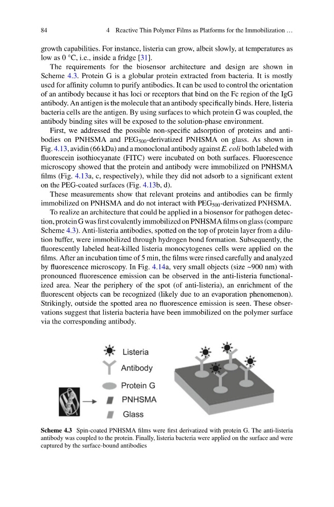
The requirements for the biosensor architecture and design are shown in
Scheme 4.3. Protein G is a globular protein extracted from bacteria. It is mostly
used for affinity column to purify antibodies. It can be used to control the orientation
of an antibody because it has loci or receptors that bind on the Fc region of the IgG
antibody. An antigen is the molecule that an antibody specifically binds. Here, listeria
bacteria cells are the antigen. By using surfaces to which protein G was coupled, the
antibody binding sites will be exposed to the solution-phase environment.
First, we addressed the possible non-specific adsorption of proteins and antibodies on PNHSMA and PEG 500 -derivatized PNHSMA on glass. As shown in
Fig. 4.13, avidin (66 kDa) and a monoclonal antibody against E. coli both labeled with
fluorescein isothiocyanate (FITC) were incubated on both surfaces. Fluorescence
microscopy showed that the protein and antibody were immobilized on PNHSMA
films (Fig. 4.13a, c, respectively), while they did not adsorb to a significant extent
on the PEG-coated surfaces (Fig. 4.13b, d).
These measurements show that relevant proteins and antibodies can be firmly
immobilized on PNHSMA and do not interact with PEG 500 -derivatized PNHSMA.
To realize an architecture that could be applied in a biosensor for pathogen detection, protein G was first covalently immobilized on PNHSMA films on glass (compare
Scheme 4.3). Anti-listeria antibodies, spotted on the top of protein layer from a dilution buffer, were immobilized through hydrogen bond formation. Subsequently, the
fluorescently labeled heat-killed listeria monocytogenes cells were applied on the
films. After an incubation time of 5 min, the films were rinsed carefully and analyzed
by fluorescence microscopy. In Fig. 4.14a, very small objects (size ~900 nm) with
pronounced fluorescence emission can be observed in the anti-listeria functionalized area. Near the periphery of the spot (of anti-listeria), an enrichment of the
fluorescent objects can be recognized (likely due to an evaporation phenomenon).
Strikingly, outside the spotted area no fluorescence emission is seen. These observations suggest that listeria bacteria have been immobilized on the polymer surface
via the corresponding antibody.
Scheme 4.3 Spin-coated PNHSMA films were first derivatized with protein G. The anti-listeria
antibody was coupled to the protein. Finally, listeria bacteria were applied on the surface and were
captured by the surface-bound antibodies
4 Reactive Thin Polymer Films as Platforms for the Immobilization …
growth capabilities. For instance, listeria can grow, albeit slowly, at temperatures as
low as 0 °C, i.e., inside a fridge [31].
The requirements for the biosensor architecture and design are shown in
Scheme 4.3. Protein G is a globular protein extracted from bacteria. It is mostly
used for affinity column to purify antibodies. It can be used to control the orientation
of an antibody because it has loci or receptors that bind on the Fc region of the IgG
antibody. An antigen is the molecule that an antibody specifically binds. Here, listeria
bacteria cells are the antigen. By using surfaces to which protein G was coupled, the
antibody binding sites will be exposed to the solution-phase environment.
First, we addressed the possible non-specific adsorption of proteins and antibodies on PNHSMA and PEG 500 -derivatized PNHSMA on glass. As shown in
Fig. 4.13, avidin (66 kDa) and a monoclonal antibody against E. coli both labeled with
fluorescein isothiocyanate (FITC) were incubated on both surfaces. Fluorescence
microscopy showed that the protein and antibody were immobilized on PNHSMA
films (Fig. 4.13a, c, respectively), while they did not adsorb to a significant extent
on the PEG-coated surfaces (Fig. 4.13b, d).
These measurements show that relevant proteins and antibodies can be firmly
immobilized on PNHSMA and do not interact with PEG 500 -derivatized PNHSMA.
To realize an architecture that could be applied in a biosensor for pathogen detection, protein G was first covalently immobilized on PNHSMA films on glass (compare
Scheme 4.3). Anti-listeria antibodies, spotted on the top of protein layer from a dilution buffer, were immobilized through hydrogen bond formation. Subsequently, the
fluorescently labeled heat-killed listeria monocytogenes cells were applied on the
films. After an incubation time of 5 min, the films were rinsed carefully and analyzed
by fluorescence microscopy. In Fig. 4.14a, very small objects (size ~900 nm) with
pronounced fluorescence emission can be observed in the anti-listeria functionalized area. Near the periphery of the spot (of anti-listeria), an enrichment of the
fluorescent objects can be recognized (likely due to an evaporation phenomenon).
Strikingly, outside the spotted area no fluorescence emission is seen. These observations suggest that listeria bacteria have been immobilized on the polymer surface
via the corresponding antibody.
Scheme 4.3 Spin-coated PNHSMA films were first derivatized with protein G. The anti-listeria
antibody was coupled to the protein. Finally, listeria bacteria were applied on the surface and were
captured by the surface-bound antibodies