

4.5 Toward the Detection of Pathogenic Bacteria on PNHSMA Films
85
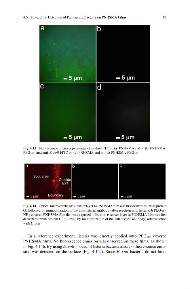
Fig. 4.13 Fluorescence microscopy images of avidin-FITC on (a) PNHSMA and on (b) PNHSMAPEG 500 , and anti-E. coli-FITC on (c) PNHSMA and on (d) PNHSMA-PEG 500
Fig. 4.14 Optical micrographs of: a sensor layer (a PNHSMA film was first derivatized with protein
G, followed by immobilization of the anti-listeria antibody) after reaction with listeria; b PEG 500 -
NH 2 covered PNHSMA film that was exposed to listeria; c sensor layer (a PNHSMA film was first
derivatized with protein G, followed by immobilization of the anti-listeria antibody) after reaction
with E. coli
In a reference experiment, listeria was directly applied onto PEG 500 covered
PNHSMA films. No fluorescence emission was observed on these films, as shown
in Fig. 4.14b. By using E. coli instead of listeria bacteria also, no fluorescence emission was detected on the surface (Fig. 4.14c). Since E. coli bacteria do not bind,
85
Fig. 4.13 Fluorescence microscopy images of avidin-FITC on (a) PNHSMA and on (b) PNHSMAPEG 500 , and anti-E. coli-FITC on (c) PNHSMA and on (d) PNHSMA-PEG 500
Fig. 4.14 Optical micrographs of: a sensor layer (a PNHSMA film was first derivatized with protein
G, followed by immobilization of the anti-listeria antibody) after reaction with listeria; b PEG 500 -
NH 2 covered PNHSMA film that was exposed to listeria; c sensor layer (a PNHSMA film was first
derivatized with protein G, followed by immobilization of the anti-listeria antibody) after reaction
with E. coli
In a reference experiment, listeria was directly applied onto PEG 500 covered
PNHSMA films. No fluorescence emission was observed on these films, as shown
in Fig. 4.14b. By using E. coli instead of listeria bacteria also, no fluorescence emission was detected on the surface (Fig. 4.14c). Since E. coli bacteria do not bind,