

4.3 Swelling Behavior and Stability of PNHSMA Films
79
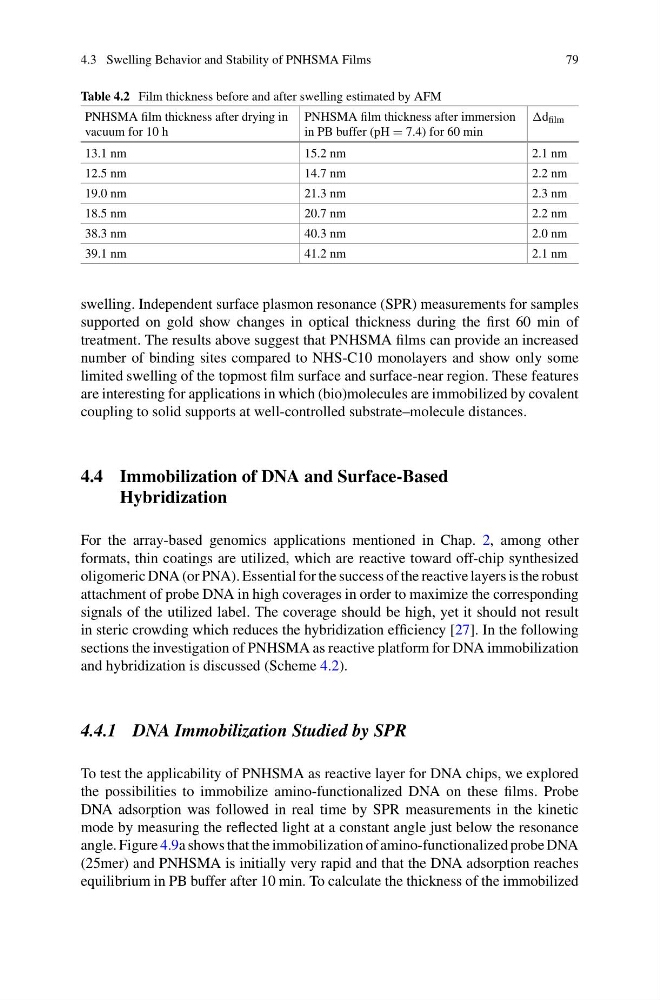
Table 4.2 Film thickness before and after swelling estimated by AFM
PNHSMA film thickness after drying in
vacuum for 10 h
PNHSMA film thickness after immersion
in PB buffer (pH = 7.4) for 60 min
d film
13.1 nm
15.2 nm
2.1 nm
12.5 nm
14.7 nm
2.2 nm
19.0 nm
21.3 nm
2.3 nm
18.5 nm
20.7 nm
2.2 nm
38.3 nm
40.3 nm
2.0 nm
39.1 nm
41.2 nm
2.1 nm
swelling. Independent surface plasmon resonance (SPR) measurements for samples
supported on gold show changes in optical thickness during the first 60 min of
treatment. The results above suggest that PNHSMA films can provide an increased
number of binding sites compared to NHS-C10 monolayers and show only some
limited swelling of the topmost film surface and surface-near region. These features
are interesting for applications in which (bio)molecules are immobilized by covalent
coupling to solid supports at well-controlled substrate–molecule distances.
4.4 Immobilization of DNA and Surface-Based
Hybridization
For the array-based genomics applications mentioned in Chap. 2, among other
formats, thin coatings are utilized, which are reactive toward off-chip synthesized
oligomeric DNA (or PNA). Essential for the success of the reactive layers is the robust
attachment of probe DNA in high coverages in order to maximize the corresponding
signals of the utilized label. The coverage should be high, yet it should not result
in steric crowding which reduces the hybridization efficiency [27]. In the following
sections the investigation of PNHSMA as reactive platform for DNA immobilization
and hybridization is discussed (Scheme 4.2).
4.4.1 DNA Immobilization Studied by SPR
To test the applicability of PNHSMA as reactive layer for DNA chips, we explored
the possibilities to immobilize amino-functionalized DNA on these films. Probe
DNA adsorption was followed in real time by SPR measurements in the kinetic
mode by measuring the reflected light at a constant angle just below the resonance
angle. Figure 4.9a shows that the immobilization of amino-functionalized probe DNA
(25mer) and PNHSMA is initially very rapid and that the DNA adsorption reaches
equilibrium in PB buffer after 10 min. To calculate the thickness of the immobilized
79
Table 4.2 Film thickness before and after swelling estimated by AFM
PNHSMA film thickness after drying in
vacuum for 10 h
PNHSMA film thickness after immersion
in PB buffer (pH = 7.4) for 60 min
d film
13.1 nm
15.2 nm
2.1 nm
12.5 nm
14.7 nm
2.2 nm
19.0 nm
21.3 nm
2.3 nm
18.5 nm
20.7 nm
2.2 nm
38.3 nm
40.3 nm
2.0 nm
39.1 nm
41.2 nm
2.1 nm
swelling. Independent surface plasmon resonance (SPR) measurements for samples
supported on gold show changes in optical thickness during the first 60 min of
treatment. The results above suggest that PNHSMA films can provide an increased
number of binding sites compared to NHS-C10 monolayers and show only some
limited swelling of the topmost film surface and surface-near region. These features
are interesting for applications in which (bio)molecules are immobilized by covalent
coupling to solid supports at well-controlled substrate–molecule distances.
4.4 Immobilization of DNA and Surface-Based
Hybridization
For the array-based genomics applications mentioned in Chap. 2, among other
formats, thin coatings are utilized, which are reactive toward off-chip synthesized
oligomeric DNA (or PNA). Essential for the success of the reactive layers is the robust
attachment of probe DNA in high coverages in order to maximize the corresponding
signals of the utilized label. The coverage should be high, yet it should not result
in steric crowding which reduces the hybridization efficiency [27]. In the following
sections the investigation of PNHSMA as reactive platform for DNA immobilization
and hybridization is discussed (Scheme 4.2).
4.4.1 DNA Immobilization Studied by SPR
To test the applicability of PNHSMA as reactive layer for DNA chips, we explored
the possibilities to immobilize amino-functionalized DNA on these films. Probe
DNA adsorption was followed in real time by SPR measurements in the kinetic
mode by measuring the reflected light at a constant angle just below the resonance
angle. Figure 4.9a shows that the immobilization of amino-functionalized probe DNA
(25mer) and PNHSMA is initially very rapid and that the DNA adsorption reaches
equilibrium in PB buffer after 10 min. To calculate the thickness of the immobilized