

4.2 Investigation of the Reactivity of PNHSMA
77
Fig. 4.6 IR spectra of NHS-C10 monolayer vs. PEG 500 -NH 2 grafted NHS-C10 SAM on gold
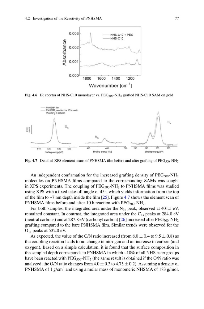
Fig. 4.7 Detailed XPS element scans of PNHSMA film before and after grafting of PEG 500 -NH 2
An independent confirmation for the increased grafting density of PEG 500 -NH 2
molecules on PNHSMA films compared to the corresponding SAMs was sought
in XPS experiments. The coupling of PEG 500 -NH 2 to PNHSMA films was studied
using XPS with a fixed take-off angle of 45°, which yields information from the top
of the film to ~7 nm depth inside the film [25]. Figure 4.7 shows the element scan of
PNHSMA films before and after 10 h reaction with PEG 500 -NH 2 .
For both samples, the integrated area under the N 1s peak, observed at 401.5 eV,
remained constant. In contrast, the integrated area under the C 1s peaks at 284.0 eV
(neutral carbons) and at 287.8 eV (carbonyl carbon) [26] increased after PEG 500 -NH 2
grafting compared to the bare PNHSMA film. Similar trends were observed for the
O 1s peaks at 532.0 eV.
As expected, the value of the C/N ratio increased (from 8.0 ± 0.4 to 9.5 ± 0.8) as
the coupling reaction leads to no change in nitrogen and an increase in carbon (and
oxygen). Based on a simple calculation, it is found that the surface composition in
the sampled depth corresponds to PNHSMA in which ~10% of all NHS ester groups
have been reacted with PEG 500 -NH 2 (the same result is obtained if the O/N ratio was
analyzed; the O/N ratio changes from 4.0 ± 0.3 to 4.75 ± 0.2). Assuming a density of
PNHSMA of 1 g/cm
3 and using a molar mass of monomeric NHSMA of 183 g/mol,
77
Fig. 4.6 IR spectra of NHS-C10 monolayer vs. PEG 500 -NH 2 grafted NHS-C10 SAM on gold
Fig. 4.7 Detailed XPS element scans of PNHSMA film before and after grafting of PEG 500 -NH 2
An independent confirmation for the increased grafting density of PEG 500 -NH 2
molecules on PNHSMA films compared to the corresponding SAMs was sought
in XPS experiments. The coupling of PEG 500 -NH 2 to PNHSMA films was studied
using XPS with a fixed take-off angle of 45°, which yields information from the top
of the film to ~7 nm depth inside the film [25]. Figure 4.7 shows the element scan of
PNHSMA films before and after 10 h reaction with PEG 500 -NH 2 .
For both samples, the integrated area under the N 1s peak, observed at 401.5 eV,
remained constant. In contrast, the integrated area under the C 1s peaks at 284.0 eV
(neutral carbons) and at 287.8 eV (carbonyl carbon) [26] increased after PEG 500 -NH 2
grafting compared to the bare PNHSMA film. Similar trends were observed for the
O 1s peaks at 532.0 eV.
As expected, the value of the C/N ratio increased (from 8.0 ± 0.4 to 9.5 ± 0.8) as
the coupling reaction leads to no change in nitrogen and an increase in carbon (and
oxygen). Based on a simple calculation, it is found that the surface composition in
the sampled depth corresponds to PNHSMA in which ~10% of all NHS ester groups
have been reacted with PEG 500 -NH 2 (the same result is obtained if the O/N ratio was
analyzed; the O/N ratio changes from 4.0 ± 0.3 to 4.75 ± 0.2). Assuming a density of
PNHSMA of 1 g/cm
3 and using a molar mass of monomeric NHSMA of 183 g/mol,