

9.3 Cell-Capture on Functionalized Hierarchical PDMS
177
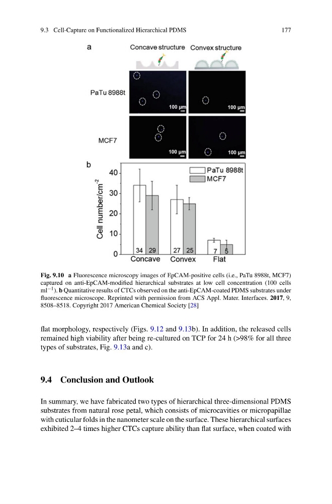
Fig. 9.10 a Fluorescence microscopy images of EpCAM-positive cells (i.e., PaTu 8988t, MCF7)
captured on anti-EpCAM-modified hierarchical substrates at low cell concentration (100 cells
ml −1 ). b Quantitative results of CTCs observed on the anti-EpCAM-coated PDMS substrates under
fluorescence microscope. Reprinted with permission from ACS Appl. Mater. Interfaces. 2017, 9,
8508–8518. Copyright 2017 American Chemical Society [28]
flat morphology, respectively (Figs. 9.12 and 9.13b). In addition, the released cells
remained high viability after being re-cultured on TCP for 24 h (>98% for all three
types of substrates, Fig. 9.13a and c).
9.4 Conclusion and Outlook
In summary, we have fabricated two types of hierarchical three-dimensional PDMS
substrates from natural rose petal, which consists of microcavities or micropapillae
with cuticular folds in the nanometer scale on the surface. These hierarchical surfaces
exhibited 2–4 times higher CTCs capture ability than flat surface, when coated with
177
Fig. 9.10 a Fluorescence microscopy images of EpCAM-positive cells (i.e., PaTu 8988t, MCF7)
captured on anti-EpCAM-modified hierarchical substrates at low cell concentration (100 cells
ml −1 ). b Quantitative results of CTCs observed on the anti-EpCAM-coated PDMS substrates under
fluorescence microscope. Reprinted with permission from ACS Appl. Mater. Interfaces. 2017, 9,
8508–8518. Copyright 2017 American Chemical Society [28]
flat morphology, respectively (Figs. 9.12 and 9.13b). In addition, the released cells
remained high viability after being re-cultured on TCP for 24 h (>98% for all three
types of substrates, Fig. 9.13a and c).
9.4 Conclusion and Outlook
In summary, we have fabricated two types of hierarchical three-dimensional PDMS
substrates from natural rose petal, which consists of microcavities or micropapillae
with cuticular folds in the nanometer scale on the surface. These hierarchical surfaces
exhibited 2–4 times higher CTCs capture ability than flat surface, when coated with