

9.3 Cell-Capture on Functionalized Hierarchical PDMS
175
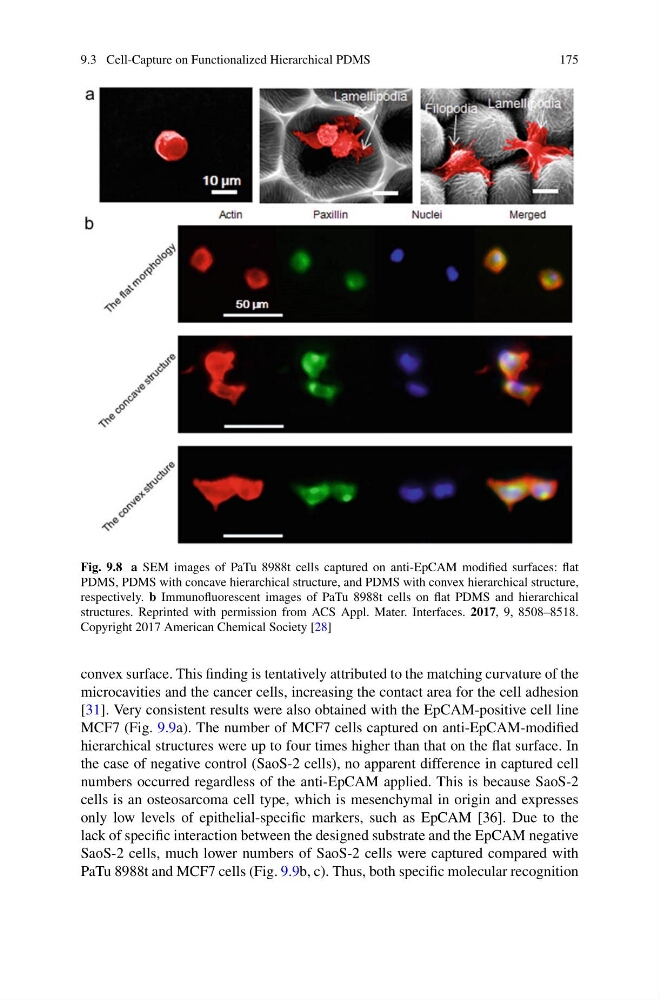
Fig. 9.8 a SEM images of PaTu 8988t cells captured on anti-EpCAM modified surfaces: flat
PDMS, PDMS with concave hierarchical structure, and PDMS with convex hierarchical structure,
respectively. b Immunofluorescent images of PaTu 8988t cells on flat PDMS and hierarchical
structures. Reprinted with permission from ACS Appl. Mater. Interfaces. 2017, 9, 8508–8518.
Copyright 2017 American Chemical Society [28]
convex surface. This finding is tentatively attributed to the matching curvature of the
microcavities and the cancer cells, increasing the contact area for the cell adhesion
[31]. Very consistent results were also obtained with the EpCAM-positive cell line
MCF7 (Fig. 9.9a). The number of MCF7 cells captured on anti-EpCAM-modified
hierarchical structures were up to four times higher than that on the flat surface. In
the case of negative control (SaoS-2 cells), no apparent difference in captured cell
numbers occurred regardless of the anti-EpCAM applied. This is because SaoS-2
cells is an osteosarcoma cell type, which is mesenchymal in origin and expresses
only low levels of epithelial-specific markers, such as EpCAM [36]. Due to the
lack of specific interaction between the designed substrate and the EpCAM negative
SaoS-2 cells, much lower numbers of SaoS-2 cells were captured compared with
PaTu 8988t and MCF7 cells (Fig. 9.9b, c). Thus, both specific molecular recognition
175
Fig. 9.8 a SEM images of PaTu 8988t cells captured on anti-EpCAM modified surfaces: flat
PDMS, PDMS with concave hierarchical structure, and PDMS with convex hierarchical structure,
respectively. b Immunofluorescent images of PaTu 8988t cells on flat PDMS and hierarchical
structures. Reprinted with permission from ACS Appl. Mater. Interfaces. 2017, 9, 8508–8518.
Copyright 2017 American Chemical Society [28]
convex surface. This finding is tentatively attributed to the matching curvature of the
microcavities and the cancer cells, increasing the contact area for the cell adhesion
[31]. Very consistent results were also obtained with the EpCAM-positive cell line
MCF7 (Fig. 9.9a). The number of MCF7 cells captured on anti-EpCAM-modified
hierarchical structures were up to four times higher than that on the flat surface. In
the case of negative control (SaoS-2 cells), no apparent difference in captured cell
numbers occurred regardless of the anti-EpCAM applied. This is because SaoS-2
cells is an osteosarcoma cell type, which is mesenchymal in origin and expresses
only low levels of epithelial-specific markers, such as EpCAM [36]. Due to the
lack of specific interaction between the designed substrate and the EpCAM negative
SaoS-2 cells, much lower numbers of SaoS-2 cells were captured compared with
PaTu 8988t and MCF7 cells (Fig. 9.9b, c). Thus, both specific molecular recognition