

9.2 Preparation of Rose Petal-Derived PDMS Surface Functionalized …
173
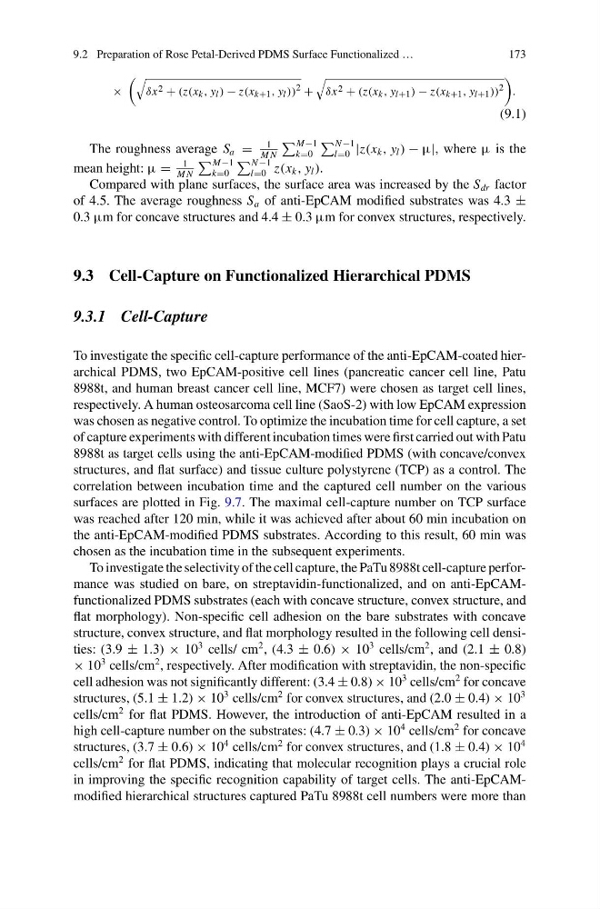
×
δx 2 + (z(x k , y l ) − z(x k+1 , y l ))
2 +
δx 2 + (z(x k , y l+1 ) − z(x k+1 , y l+1 ))
2
.
(9.1)
The roughness average S a =
1
M N
M−1
k=0
N −1
l=0 |z(x k , y l ) − μ|, where μ is the
mean height: μ =
1
M N
M−1
k=0
N −1
l=0 z(x k , y l ).
Compared with plane surfaces, the surface area was increased by the S dr factor
of 4.5. The average roughness S a of anti-EpCAM modified substrates was 4.3 ±
0.3 μm for concave structures and 4.4 ± 0.3 μm for convex structures, respectively.
9.3 Cell-Capture on Functionalized Hierarchical PDMS
9.3.1 Cell-Capture
To investigate the specific cell-capture performance of the anti-EpCAM-coated hierarchical PDMS, two EpCAM-positive cell lines (pancreatic cancer cell line, Patu
8988t, and human breast cancer cell line, MCF7) were chosen as target cell lines,
respectively. A human osteosarcoma cell line (SaoS-2) with low EpCAM expression
was chosen as negative control. To optimize the incubation time for cell capture, a set
of capture experiments with different incubation times were first carried out with Patu
8988t as target cells using the anti-EpCAM-modified PDMS (with concave/convex
structures, and flat surface) and tissue culture polystyrene (TCP) as a control. The
correlation between incubation time and the captured cell number on the various
surfaces are plotted in Fig. 9.7. The maximal cell-capture number on TCP surface
was reached after 120 min, while it was achieved after about 60 min incubation on
the anti-EpCAM-modified PDMS substrates. According to this result, 60 min was
chosen as the incubation time in the subsequent experiments.
To investigate the selectivity of the cell capture, the PaTu 8988t cell-capture performance was studied on bare, on streptavidin-functionalized, and on anti-EpCAMfunctionalized PDMS substrates (each with concave structure, convex structure, and
flat morphology). Non-specific cell adhesion on the bare substrates with concave
structure, convex structure, and flat morphology resulted in the following cell densities: (3.9 ± 1.3) × 10
3 cells/ cm
2 , (4.3 ± 0.6) × 10
3 cells/cm
2 , and (2.1 ± 0.8)
× 10
3 cells/cm
2 , respectively. After modification with streptavidin, the non-specific
cell adhesion was not significantly different: (3.4 ± 0.8) × 10
3 cells/cm
2 for concave
structures, (5.1 ± 1.2) × 10
3 cells/cm
2 for convex structures, and (2.0 ± 0.4) × 10
3
cells/cm
2 for flat PDMS. However, the introduction of anti-EpCAM resulted in a
high cell-capture number on the substrates: (4.7 ± 0.3) × 10
4 cells/cm
2 for concave
structures, (3.7 ± 0.6) × 10
4 cells/cm
2 for convex structures, and (1.8 ± 0.4) × 10
4
cells/cm
2 for flat PDMS, indicating that molecular recognition plays a crucial role
in improving the specific recognition capability of target cells. The anti-EpCAMmodified hierarchical structures captured PaTu 8988t cell numbers were more than
173
×
δx 2 + (z(x k , y l ) − z(x k+1 , y l ))
2 +
δx 2 + (z(x k , y l+1 ) − z(x k+1 , y l+1 ))
2
.
(9.1)
The roughness average S a =
1
M N
M−1
k=0
N −1
l=0 |z(x k , y l ) − μ|, where μ is the
mean height: μ =
1
M N
M−1
k=0
N −1
l=0 z(x k , y l ).
Compared with plane surfaces, the surface area was increased by the S dr factor
of 4.5. The average roughness S a of anti-EpCAM modified substrates was 4.3 ±
0.3 μm for concave structures and 4.4 ± 0.3 μm for convex structures, respectively.
9.3 Cell-Capture on Functionalized Hierarchical PDMS
9.3.1 Cell-Capture
To investigate the specific cell-capture performance of the anti-EpCAM-coated hierarchical PDMS, two EpCAM-positive cell lines (pancreatic cancer cell line, Patu
8988t, and human breast cancer cell line, MCF7) were chosen as target cell lines,
respectively. A human osteosarcoma cell line (SaoS-2) with low EpCAM expression
was chosen as negative control. To optimize the incubation time for cell capture, a set
of capture experiments with different incubation times were first carried out with Patu
8988t as target cells using the anti-EpCAM-modified PDMS (with concave/convex
structures, and flat surface) and tissue culture polystyrene (TCP) as a control. The
correlation between incubation time and the captured cell number on the various
surfaces are plotted in Fig. 9.7. The maximal cell-capture number on TCP surface
was reached after 120 min, while it was achieved after about 60 min incubation on
the anti-EpCAM-modified PDMS substrates. According to this result, 60 min was
chosen as the incubation time in the subsequent experiments.
To investigate the selectivity of the cell capture, the PaTu 8988t cell-capture performance was studied on bare, on streptavidin-functionalized, and on anti-EpCAMfunctionalized PDMS substrates (each with concave structure, convex structure, and
flat morphology). Non-specific cell adhesion on the bare substrates with concave
structure, convex structure, and flat morphology resulted in the following cell densities: (3.9 ± 1.3) × 10
3 cells/ cm
2 , (4.3 ± 0.6) × 10
3 cells/cm
2 , and (2.1 ± 0.8)
× 10
3 cells/cm
2 , respectively. After modification with streptavidin, the non-specific
cell adhesion was not significantly different: (3.4 ± 0.8) × 10
3 cells/cm
2 for concave
structures, (5.1 ± 1.2) × 10
3 cells/cm
2 for convex structures, and (2.0 ± 0.4) × 10
3
cells/cm
2 for flat PDMS. However, the introduction of anti-EpCAM resulted in a
high cell-capture number on the substrates: (4.7 ± 0.3) × 10
4 cells/cm
2 for concave
structures, (3.7 ± 0.6) × 10
4 cells/cm
2 for convex structures, and (1.8 ± 0.4) × 10
4
cells/cm
2 for flat PDMS, indicating that molecular recognition plays a crucial role
in improving the specific recognition capability of target cells. The anti-EpCAMmodified hierarchical structures captured PaTu 8988t cell numbers were more than