

142
7 Reactive μCP on Ultrathin Block Copolymer Films …
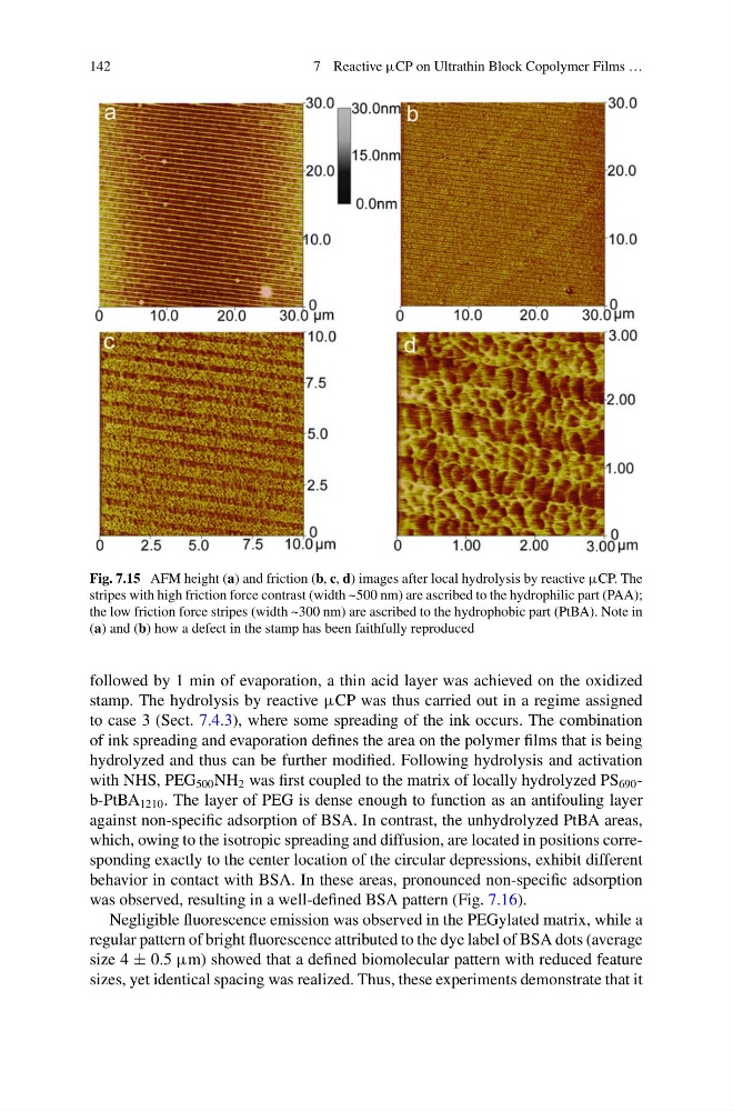
Fig. 7.15 AFM height (a) and friction (b, c, d) images after local hydrolysis by reactive μCP. The
stripes with high friction force contrast (width ~500 nm) are ascribed to the hydrophilic part (PAA);
the low friction force stripes (width ~300 nm) are ascribed to the hydrophobic part (PtBA). Note in
(a) and (b) how a defect in the stamp has been faithfully reproduced
followed by 1 min of evaporation, a thin acid layer was achieved on the oxidized
stamp. The hydrolysis by reactive μCP was thus carried out in a regime assigned
to case 3 (Sect. 7.4.3), where some spreading of the ink occurs. The combination
of ink spreading and evaporation defines the area on the polymer films that is being
hydrolyzed and thus can be further modified. Following hydrolysis and activation
with NHS, PEG 500 NH 2 was first coupled to the matrix of locally hydrolyzed PS 690 -
b-PtBA 1210 . The layer of PEG is dense enough to function as an antifouling layer
against non-specific adsorption of BSA. In contrast, the unhydrolyzed PtBA areas,
which, owing to the isotropic spreading and diffusion, are located in positions corresponding exactly to the center location of the circular depressions, exhibit different
behavior in contact with BSA. In these areas, pronounced non-specific adsorption
was observed, resulting in a well-defined BSA pattern (Fig. 7.16).
Negligible fluorescence emission was observed in the PEGylated matrix, while a
regular pattern of bright fluorescence attributed to the dye label of BSA dots (average
size 4 ± 0.5 μm) showed that a defined biomolecular pattern with reduced feature
sizes, yet identical spacing was realized. Thus, these experiments demonstrate that it
7 Reactive μCP on Ultrathin Block Copolymer Films …
Fig. 7.15 AFM height (a) and friction (b, c, d) images after local hydrolysis by reactive μCP. The
stripes with high friction force contrast (width ~500 nm) are ascribed to the hydrophilic part (PAA);
the low friction force stripes (width ~300 nm) are ascribed to the hydrophobic part (PtBA). Note in
(a) and (b) how a defect in the stamp has been faithfully reproduced
followed by 1 min of evaporation, a thin acid layer was achieved on the oxidized
stamp. The hydrolysis by reactive μCP was thus carried out in a regime assigned
to case 3 (Sect. 7.4.3), where some spreading of the ink occurs. The combination
of ink spreading and evaporation defines the area on the polymer films that is being
hydrolyzed and thus can be further modified. Following hydrolysis and activation
with NHS, PEG 500 NH 2 was first coupled to the matrix of locally hydrolyzed PS 690 -
b-PtBA 1210 . The layer of PEG is dense enough to function as an antifouling layer
against non-specific adsorption of BSA. In contrast, the unhydrolyzed PtBA areas,
which, owing to the isotropic spreading and diffusion, are located in positions corresponding exactly to the center location of the circular depressions, exhibit different
behavior in contact with BSA. In these areas, pronounced non-specific adsorption
was observed, resulting in a well-defined BSA pattern (Fig. 7.16).
Negligible fluorescence emission was observed in the PEGylated matrix, while a
regular pattern of bright fluorescence attributed to the dye label of BSA dots (average
size 4 ± 0.5 μm) showed that a defined biomolecular pattern with reduced feature
sizes, yet identical spacing was realized. Thus, these experiments demonstrate that it