

118
6 Fabrication of Robust Biomolecular Patterns …
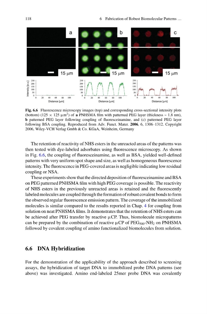
Fig. 6.6 Fluorescence microscopy images (top) and corresponding cross-sectional intensity plots
(bottom) (125 × 125 μm 2 ) of a PNHSMA film with patterned PEG layer (thickness ~ 1.8 nm),
b patterned PEG layer following coupling of fluoresceinamine, and (c) patterned PEG layer
following BSA coupling. Reproduced from Adv. Funct. Mater. 2006, 6, 1306–1312. Copyright
2006, Wiley-VCH Verlag Gmbh & Co. KGaA, Weinheim, Germany
The retention of reactivity of NHS esters in the unreacted areas of the patterns was
then tested with dye-labeled adsorbates using fluorescence microscopy. As shown
in Fig. 6.6, the coupling of fluoresceinamine, as well as BSA, yielded well-defined
patterns with very uniform spot shape and size, as well as homogeneous fluorescence
intensity. The fluorescence in PEG-covered areas is negligible indicating low residual
coupling or NSA.
These experiments show that the directed deposition of fluoresceinamine and BSA
on PEG patterned PNHSMA film with high PEG coverage is possible. The reactivity
of NHS esters in the previously unreacted areas is retained and the fluorescently
labeled molecules are coupled through the formation of robust covalent bonds to form
the observed regular fluorescence emission pattern. The coverage of the immobilized
molecules is similar compared to the results reported in Chap. 4 for coupling from
solution on neat PNHSMA films. It demonstrates that the retention of NHS esters can
be achieved after PEG transfer by reactive μCP. Thus, biomolecule micropatterns
can be prepared by the combination of reactive μCP of PEG 500 -NH 2 on PNHSMA
followed by covalent coupling of amino functionalized biomolecules from solution.
6.6 DNA Hybridization
For the demonstration of the applicability of the approach described to screening
assays, the hybridization of target DNA to immobilized probe DNA patterns (see
above) was investigated. Amino end-labeled 25mer probe DNA was covalently
6 Fabrication of Robust Biomolecular Patterns …
Fig. 6.6 Fluorescence microscopy images (top) and corresponding cross-sectional intensity plots
(bottom) (125 × 125 μm 2 ) of a PNHSMA film with patterned PEG layer (thickness ~ 1.8 nm),
b patterned PEG layer following coupling of fluoresceinamine, and (c) patterned PEG layer
following BSA coupling. Reproduced from Adv. Funct. Mater. 2006, 6, 1306–1312. Copyright
2006, Wiley-VCH Verlag Gmbh & Co. KGaA, Weinheim, Germany
The retention of reactivity of NHS esters in the unreacted areas of the patterns was
then tested with dye-labeled adsorbates using fluorescence microscopy. As shown
in Fig. 6.6, the coupling of fluoresceinamine, as well as BSA, yielded well-defined
patterns with very uniform spot shape and size, as well as homogeneous fluorescence
intensity. The fluorescence in PEG-covered areas is negligible indicating low residual
coupling or NSA.
These experiments show that the directed deposition of fluoresceinamine and BSA
on PEG patterned PNHSMA film with high PEG coverage is possible. The reactivity
of NHS esters in the previously unreacted areas is retained and the fluorescently
labeled molecules are coupled through the formation of robust covalent bonds to form
the observed regular fluorescence emission pattern. The coverage of the immobilized
molecules is similar compared to the results reported in Chap. 4 for coupling from
solution on neat PNHSMA films. It demonstrates that the retention of NHS esters can
be achieved after PEG transfer by reactive μCP. Thus, biomolecule micropatterns
can be prepared by the combination of reactive μCP of PEG 500 -NH 2 on PNHSMA
followed by covalent coupling of amino functionalized biomolecules from solution.
6.6 DNA Hybridization
For the demonstration of the applicability of the approach described to screening
assays, the hybridization of target DNA to immobilized probe DNA patterns (see
above) was investigated. Amino end-labeled 25mer probe DNA was covalently