

6.6 DNA Hybridization
119
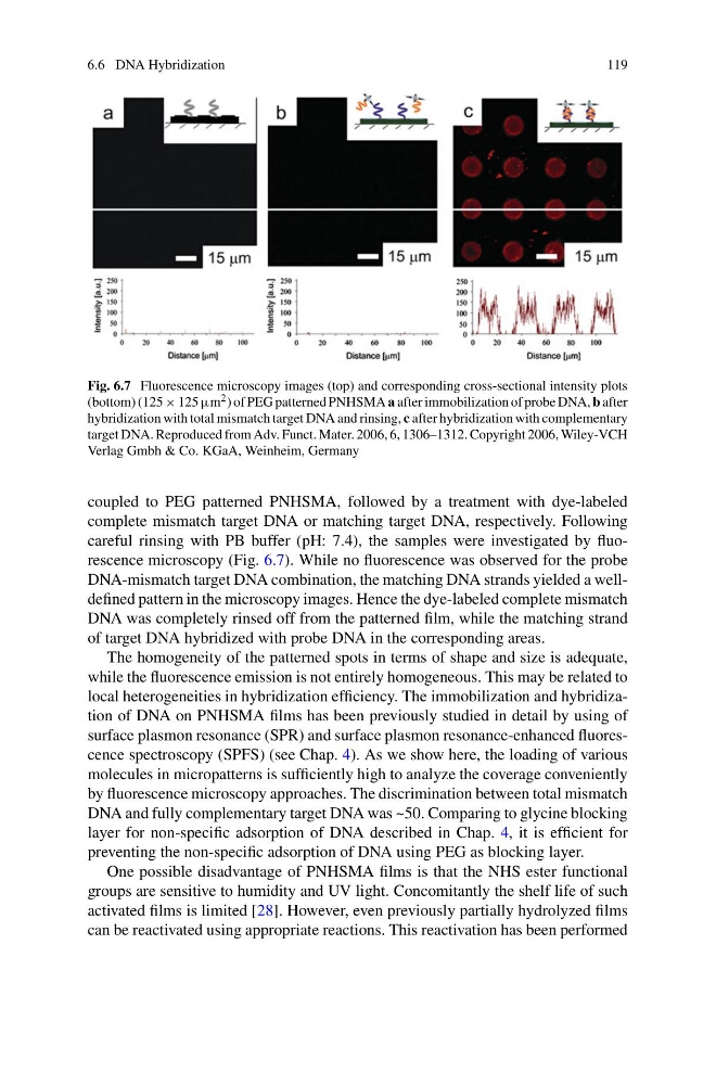
Fig. 6.7 Fluorescence microscopy images (top) and corresponding cross-sectional intensity plots
(bottom) (125 × 125 μm 2 ) of PEG patterned PNHSMA a after immobilization of probe DNA, b after
hybridization with total mismatch target DNA and rinsing, c after hybridization with complementary
target DNA. Reproduced from Adv. Funct. Mater. 2006, 6, 1306–1312. Copyright 2006, Wiley-VCH
Verlag Gmbh & Co. KGaA, Weinheim, Germany
coupled to PEG patterned PNHSMA, followed by a treatment with dye-labeled
complete mismatch target DNA or matching target DNA, respectively. Following
careful rinsing with PB buffer (pH: 7.4), the samples were investigated by fluorescence microscopy (Fig. 6.7). While no fluorescence was observed for the probe
DNA-mismatch target DNA combination, the matching DNA strands yielded a welldefined pattern in the microscopy images. Hence the dye-labeled complete mismatch
DNA was completely rinsed off from the patterned film, while the matching strand
of target DNA hybridized with probe DNA in the corresponding areas.
The homogeneity of the patterned spots in terms of shape and size is adequate,
while the fluorescence emission is not entirely homogeneous. This may be related to
local heterogeneities in hybridization efficiency. The immobilization and hybridization of DNA on PNHSMA films has been previously studied in detail by using of
surface plasmon resonance (SPR) and surface plasmon resonance-enhanced fluorescence spectroscopy (SPFS) (see Chap. 4). As we show here, the loading of various
molecules in micropatterns is sufficiently high to analyze the coverage conveniently
by fluorescence microscopy approaches. The discrimination between total mismatch
DNA and fully complementary target DNA was ~50. Comparing to glycine blocking
layer for non-specific adsorption of DNA described in Chap. 4, it is efficient for
preventing the non-specific adsorption of DNA using PEG as blocking layer.
One possible disadvantage of PNHSMA films is that the NHS ester functional
groups are sensitive to humidity and UV light. Concomitantly the shelf life of such
activated films is limited [28]. However, even previously partially hydrolyzed films
can be reactivated using appropriate reactions. This reactivation has been performed
119
Fig. 6.7 Fluorescence microscopy images (top) and corresponding cross-sectional intensity plots
(bottom) (125 × 125 μm 2 ) of PEG patterned PNHSMA a after immobilization of probe DNA, b after
hybridization with total mismatch target DNA and rinsing, c after hybridization with complementary
target DNA. Reproduced from Adv. Funct. Mater. 2006, 6, 1306–1312. Copyright 2006, Wiley-VCH
Verlag Gmbh & Co. KGaA, Weinheim, Germany
coupled to PEG patterned PNHSMA, followed by a treatment with dye-labeled
complete mismatch target DNA or matching target DNA, respectively. Following
careful rinsing with PB buffer (pH: 7.4), the samples were investigated by fluorescence microscopy (Fig. 6.7). While no fluorescence was observed for the probe
DNA-mismatch target DNA combination, the matching DNA strands yielded a welldefined pattern in the microscopy images. Hence the dye-labeled complete mismatch
DNA was completely rinsed off from the patterned film, while the matching strand
of target DNA hybridized with probe DNA in the corresponding areas.
The homogeneity of the patterned spots in terms of shape and size is adequate,
while the fluorescence emission is not entirely homogeneous. This may be related to
local heterogeneities in hybridization efficiency. The immobilization and hybridization of DNA on PNHSMA films has been previously studied in detail by using of
surface plasmon resonance (SPR) and surface plasmon resonance-enhanced fluorescence spectroscopy (SPFS) (see Chap. 4). As we show here, the loading of various
molecules in micropatterns is sufficiently high to analyze the coverage conveniently
by fluorescence microscopy approaches. The discrimination between total mismatch
DNA and fully complementary target DNA was ~50. Comparing to glycine blocking
layer for non-specific adsorption of DNA described in Chap. 4, it is efficient for
preventing the non-specific adsorption of DNA using PEG as blocking layer.
One possible disadvantage of PNHSMA films is that the NHS ester functional
groups are sensitive to humidity and UV light. Concomitantly the shelf life of such
activated films is limited [28]. However, even previously partially hydrolyzed films
can be reactivated using appropriate reactions. This reactivation has been performed