

5.4 Cell Adhesion Studies on PS 690 -b-PtBA 1210 Films
103
Especially, the cell surface coverage was found to decrease very much and only very
few isolated cells were found on the PEG 500 -NH 2 functionalized surface as seen in
Fig. 5.9c. The result demonstrates that PEG 500 -NH 2 functionalized surfaces can be
used as antifouling layer for preventing the cell interaction with the substrate.
Subsequently, the interactions of pancreatic cancer cells with PEG-functionalized
PS 690 -b-PtBA 1210 films were investigated in collaboration with Dr. J. Schnekenburger
from the University of Münster in Germany. In the attempt to optimize the blocking
ability of PEG layers to prevent adhesion of the pancreatic cancer cells, two different
types of PEG molecules with different weight average molar mass M n were used,
that is, PEG 500 -NH 2 and PEG 5000 -NH 2 . The rational behind this experiment is that
the chain length and grafting density of PEG molecules exert a large influence on
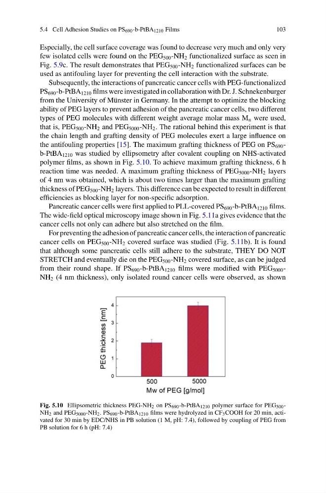
the antifouling properties [15]. The maximum grafting thickness of PEG on PS 690 -
b-PtBA 1210 was studied by ellipsometry after covalent coupling on NHS-activated
polymer films, as shown in Fig. 5.10. To achieve maximum grafting thickness, 6 h
reaction time was needed. A maximum grafting thickness of PEG 5000 -NH 2 layers
of 4 nm was obtained, which is about two times larger than the maximum grafting
thickness of PEG 500 -NH 2 layers. This difference can be expected to result in different
efficiencies as blocking layer for non-specific adsorption.
Pancreatic cancer cells were first applied to PLL-covered PS 690 -b-PtBA 1210 films.
The wide-field optical microscopy image shown in Fig. 5.11a gives evidence that the
cancer cells not only can adhere but also stretched on the film.
For preventing the adhesion of pancreatic cancer cells, the interaction of pancreatic
cancer cells on PEG 500 -NH 2 covered surface was studied (Fig. 5.11b). It is found
that although some pancreatic cells still adhere to the substrate, THEY DO NOT
STRETCH and eventually die on the PEG 500 -NH 2 covered surface, as can be judged
from their round shape. If PS 690 -b-PtBA 1210 films were modified with PEG 5000 -
NH 2 (4 nm thickness), only isolated round cancer cells were observed, as shown
Fig. 5.10 Ellipsometric thickness PEG-NH 2 on PS 690 -b-PtBA 1210 polymer surface for PEG 500 -
NH 2 and PEG 5000 -NH 2 . PS 690 -b-PtBA 1210 films were hydrolyzed in CF 3 COOH for 20 min, activated for 30 min by EDC/NHS in PB solution (1 M, pH: 7.4), followed by coupling of PEG from
PB solution for 6 h (pH: 7.4)
103
Especially, the cell surface coverage was found to decrease very much and only very
few isolated cells were found on the PEG 500 -NH 2 functionalized surface as seen in
Fig. 5.9c. The result demonstrates that PEG 500 -NH 2 functionalized surfaces can be
used as antifouling layer for preventing the cell interaction with the substrate.
Subsequently, the interactions of pancreatic cancer cells with PEG-functionalized
PS 690 -b-PtBA 1210 films were investigated in collaboration with Dr. J. Schnekenburger
from the University of Münster in Germany. In the attempt to optimize the blocking
ability of PEG layers to prevent adhesion of the pancreatic cancer cells, two different
types of PEG molecules with different weight average molar mass M n were used,
that is, PEG 500 -NH 2 and PEG 5000 -NH 2 . The rational behind this experiment is that
the chain length and grafting density of PEG molecules exert a large influence on
the antifouling properties [15]. The maximum grafting thickness of PEG on PS 690 -
b-PtBA 1210 was studied by ellipsometry after covalent coupling on NHS-activated
polymer films, as shown in Fig. 5.10. To achieve maximum grafting thickness, 6 h
reaction time was needed. A maximum grafting thickness of PEG 5000 -NH 2 layers
of 4 nm was obtained, which is about two times larger than the maximum grafting
thickness of PEG 500 -NH 2 layers. This difference can be expected to result in different
efficiencies as blocking layer for non-specific adsorption.
Pancreatic cancer cells were first applied to PLL-covered PS 690 -b-PtBA 1210 films.
The wide-field optical microscopy image shown in Fig. 5.11a gives evidence that the
cancer cells not only can adhere but also stretched on the film.
For preventing the adhesion of pancreatic cancer cells, the interaction of pancreatic
cancer cells on PEG 500 -NH 2 covered surface was studied (Fig. 5.11b). It is found
that although some pancreatic cells still adhere to the substrate, THEY DO NOT
STRETCH and eventually die on the PEG 500 -NH 2 covered surface, as can be judged
from their round shape. If PS 690 -b-PtBA 1210 films were modified with PEG 5000 -
NH 2 (4 nm thickness), only isolated round cancer cells were observed, as shown
Fig. 5.10 Ellipsometric thickness PEG-NH 2 on PS 690 -b-PtBA 1210 polymer surface for PEG 500 -
NH 2 and PEG 5000 -NH 2 . PS 690 -b-PtBA 1210 films were hydrolyzed in CF 3 COOH for 20 min, activated for 30 min by EDC/NHS in PB solution (1 M, pH: 7.4), followed by coupling of PEG from
PB solution for 6 h (pH: 7.4)