

5.3 Covalent Coupling of Biomolecules to Activated PS 690 -b-PtBA 1210 Films
101
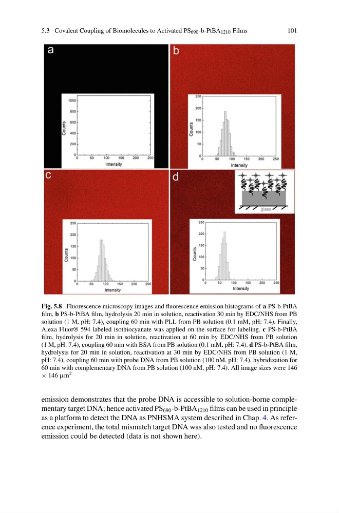
Fig. 5.8 Fluorescence microscopy images and fluorescence emission histograms of a PS-b-PtBA
film, b PS-b-PtBA film, hydrolysis 20 min in solution, reactivation 30 min by EDC/NHS from PB
solution (1 M, pH: 7.4), coupling 60 min with PLL from PB solution (0.1 mM, pH: 7.4). Finally,
Alexa Fluor® 594 labeled isothiocyanate was applied on the surface for labeling. c PS-b-PtBA
film, hydrolysis for 20 min in solution, reactivation at 60 min by EDC/NHS from PB solution
(1 M, pH: 7.4), coupling 60 min with BSA from PB solution (0.1 mM, pH: 7.4). d PS-b-PtBA film,
hydrolysis for 20 min in solution, reactivation at 30 min by EDC/NHS from PB solution (1 M,
pH: 7.4), coupling 60 min with probe DNA from PB solution (100 nM, pH: 7.4), hybridization for
60 min with complementary DNA from PB solution (100 nM, pH: 7.4). All image sizes were 146
× 146 μm 2
emission demonstrates that the probe DNA is accessible to solution-borne complementary target DNA; hence activated PS 690 -b-PtBA 1210 films can be used in principle
as a platform to detect the DNA as PNHSMA system described in Chap. 4. As reference experiment, the total mismatch target DNA was also tested and no fluorescence
emission could be detected (data is not shown here).
101
Fig. 5.8 Fluorescence microscopy images and fluorescence emission histograms of a PS-b-PtBA
film, b PS-b-PtBA film, hydrolysis 20 min in solution, reactivation 30 min by EDC/NHS from PB
solution (1 M, pH: 7.4), coupling 60 min with PLL from PB solution (0.1 mM, pH: 7.4). Finally,
Alexa Fluor® 594 labeled isothiocyanate was applied on the surface for labeling. c PS-b-PtBA
film, hydrolysis for 20 min in solution, reactivation at 60 min by EDC/NHS from PB solution
(1 M, pH: 7.4), coupling 60 min with BSA from PB solution (0.1 mM, pH: 7.4). d PS-b-PtBA film,
hydrolysis for 20 min in solution, reactivation at 30 min by EDC/NHS from PB solution (1 M,
pH: 7.4), coupling 60 min with probe DNA from PB solution (100 nM, pH: 7.4), hybridization for
60 min with complementary DNA from PB solution (100 nM, pH: 7.4). All image sizes were 146
× 146 μm 2
emission demonstrates that the probe DNA is accessible to solution-borne complementary target DNA; hence activated PS 690 -b-PtBA 1210 films can be used in principle
as a platform to detect the DNA as PNHSMA system described in Chap. 4. As reference experiment, the total mismatch target DNA was also tested and no fluorescence
emission could be detected (data is not shown here).