

80
3 In-Tether Chiral Center Induced Helical Peptide Modulators …
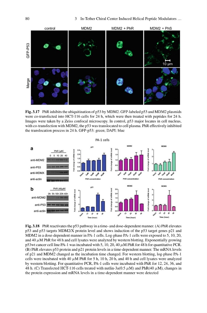
Fig. 3.17 PhR inhibits the ubiquitination of p53 by MDM2. GFP-labeled p53 and MDM2 plasmids
were co-transfected into HCT-116 cells for 24 h, which were then treated with peptides for 24 h.
Images were taken by a Zeiss confocal microscopy. In control, p53 major locates in cell nucleus,
with co-transfection with MDM2, the p53 was translocated to cell plasma. PhR effectively inhibited
the translocation process in 24 h. GFP-p53: green; DAPI: blue
Fig. 3.18 PhR reactivates the p53 pathway in a time- and dose-dependent manner. (A) PhR elevates
p53 and p53 targets MDM2/X protein level and shows induction of the p53 target genes p21 and
MDM2 in a dose-dependent manner in PA-1 cells. Log-phase PA-1 cells were exposed to 5, 10, 20,
and 40 µM PhR for 48 h and cell lysates were analyzed by western blotting. Exponentially growing
p53wt cancer cell line PA-1 was incubated with 5, 10, 20, 40 µM PhR for 48 h for quantitative PCR.
(B) PhR elevates p53 protein and p21 protein levels in a time-dependent manner. The mRNA levels
of p21 and MDM2 changed as the incubation time changed. For western blotting, log-phase PA-1
cells were incubated with 40 µM PhR for 5 h, 10 h, 20 h, and 40 h and cell lysates were analyzed
by western blotting. For quantitative PCR, PA-1 cells were incubated with PhR for 12, 24, 36, and
48 h. (C) Transfected HCT-116 cells treated with nutlin-3a(0.5 µM) and PhR(40 µM), changes in
the protein expression and mRNA levels in a time-dependent manner were detected
3 In-Tether Chiral Center Induced Helical Peptide Modulators …
Fig. 3.17 PhR inhibits the ubiquitination of p53 by MDM2. GFP-labeled p53 and MDM2 plasmids
were co-transfected into HCT-116 cells for 24 h, which were then treated with peptides for 24 h.
Images were taken by a Zeiss confocal microscopy. In control, p53 major locates in cell nucleus,
with co-transfection with MDM2, the p53 was translocated to cell plasma. PhR effectively inhibited
the translocation process in 24 h. GFP-p53: green; DAPI: blue
Fig. 3.18 PhR reactivates the p53 pathway in a time- and dose-dependent manner. (A) PhR elevates
p53 and p53 targets MDM2/X protein level and shows induction of the p53 target genes p21 and
MDM2 in a dose-dependent manner in PA-1 cells. Log-phase PA-1 cells were exposed to 5, 10, 20,
and 40 µM PhR for 48 h and cell lysates were analyzed by western blotting. Exponentially growing
p53wt cancer cell line PA-1 was incubated with 5, 10, 20, 40 µM PhR for 48 h for quantitative PCR.
(B) PhR elevates p53 protein and p21 protein levels in a time-dependent manner. The mRNA levels
of p21 and MDM2 changed as the incubation time changed. For western blotting, log-phase PA-1
cells were incubated with 40 µM PhR for 5 h, 10 h, 20 h, and 40 h and cell lysates were analyzed
by western blotting. For quantitative PCR, PA-1 cells were incubated with PhR for 12, 24, 36, and
48 h. (C) Transfected HCT-116 cells treated with nutlin-3a(0.5 µM) and PhR(40 µM), changes in
the protein expression and mRNA levels in a time-dependent manner were detected