

3.2 Results
79
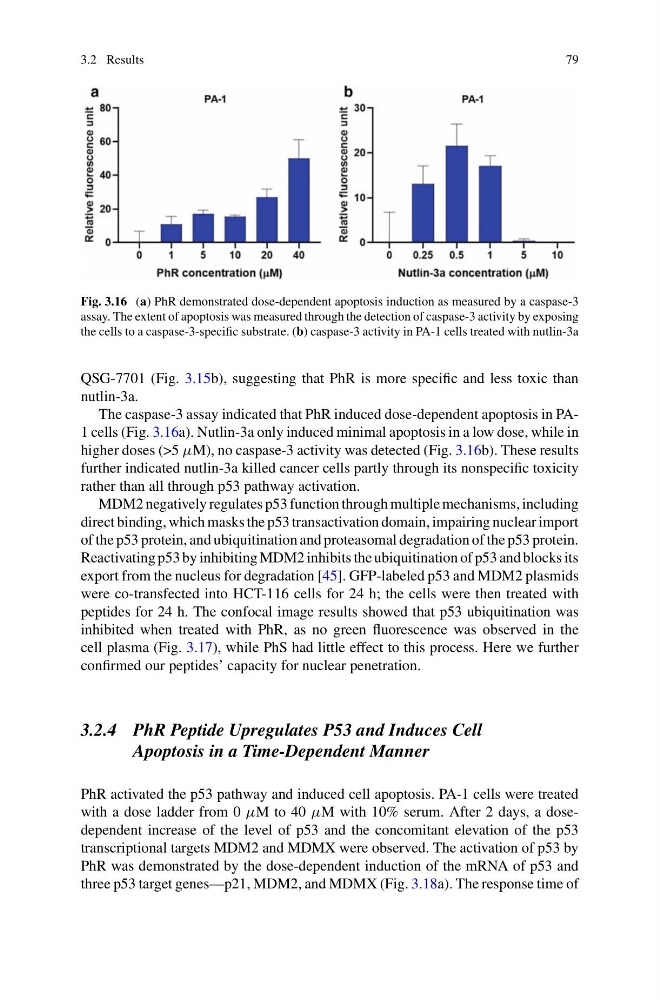
Fig. 3.16 (a) PhR demonstrated dose-dependent apoptosis induction as measured by a caspase-3
assay. The extent of apoptosis was measured through the detection of caspase-3 activity by exposing
the cells to a caspase-3-specific substrate. (b) caspase-3 activity in PA-1 cells treated with nutlin-3a
QSG-7701 (Fig. 3.15b), suggesting that PhR is more specific and less toxic than
nutlin-3a.
The caspase-3 assay indicated that PhR induced dose-dependent apoptosis in PA1 cells (Fig. 3.16a). Nutlin-3a only induced minimal apoptosis in a low dose, while in
higher doses (>5 µM), no caspase-3 activity was detected (Fig. 3.16b). These results
further indicated nutlin-3a killed cancer cells partly through its nonspecific toxicity
rather than all through p53 pathway activation.
MDM2 negatively regulates p53 function through multiple mechanisms, including
direct binding, which masks the p53 transactivation domain, impairing nuclear import
of the p53 protein, and ubiquitination and proteasomal degradation of the p53 protein.
Reactivating p53 by inhibiting MDM2 inhibits the ubiquitination of p53 and blocks its
export from the nucleus for degradation [45]. GFP-labeled p53 and MDM2 plasmids
were co-transfected into HCT-116 cells for 24 h; the cells were then treated with
peptides for 24 h. The confocal image results showed that p53 ubiquitination was
inhibited when treated with PhR, as no green fluorescence was observed in the
cell plasma (Fig. 3.17), while PhS had little effect to this process. Here we further
confirmed our peptides’ capacity for nuclear penetration.
3.2.4 PhR Peptide Upregulates P53 and Induces Cell
Apoptosis in a Time-Dependent Manner
PhR activated the p53 pathway and induced cell apoptosis. PA-1 cells were treated
with a dose ladder from 0 µM to 40 µM with 10% serum. After 2 days, a dosedependent increase of the level of p53 and the concomitant elevation of the p53
transcriptional targets MDM2 and MDMX were observed. The activation of p53 by
PhR was demonstrated by the dose-dependent induction of the mRNA of p53 and
three p53 target genes—p21, MDM2, and MDMX (Fig. 3.18a). The response time of
79
Fig. 3.16 (a) PhR demonstrated dose-dependent apoptosis induction as measured by a caspase-3
assay. The extent of apoptosis was measured through the detection of caspase-3 activity by exposing
the cells to a caspase-3-specific substrate. (b) caspase-3 activity in PA-1 cells treated with nutlin-3a
QSG-7701 (Fig. 3.15b), suggesting that PhR is more specific and less toxic than
nutlin-3a.
The caspase-3 assay indicated that PhR induced dose-dependent apoptosis in PA1 cells (Fig. 3.16a). Nutlin-3a only induced minimal apoptosis in a low dose, while in
higher doses (>5 µM), no caspase-3 activity was detected (Fig. 3.16b). These results
further indicated nutlin-3a killed cancer cells partly through its nonspecific toxicity
rather than all through p53 pathway activation.
MDM2 negatively regulates p53 function through multiple mechanisms, including
direct binding, which masks the p53 transactivation domain, impairing nuclear import
of the p53 protein, and ubiquitination and proteasomal degradation of the p53 protein.
Reactivating p53 by inhibiting MDM2 inhibits the ubiquitination of p53 and blocks its
export from the nucleus for degradation [45]. GFP-labeled p53 and MDM2 plasmids
were co-transfected into HCT-116 cells for 24 h; the cells were then treated with
peptides for 24 h. The confocal image results showed that p53 ubiquitination was
inhibited when treated with PhR, as no green fluorescence was observed in the
cell plasma (Fig. 3.17), while PhS had little effect to this process. Here we further
confirmed our peptides’ capacity for nuclear penetration.
3.2.4 PhR Peptide Upregulates P53 and Induces Cell
Apoptosis in a Time-Dependent Manner
PhR activated the p53 pathway and induced cell apoptosis. PA-1 cells were treated
with a dose ladder from 0 µM to 40 µM with 10% serum. After 2 days, a dosedependent increase of the level of p53 and the concomitant elevation of the p53
transcriptional targets MDM2 and MDMX were observed. The activation of p53 by
PhR was demonstrated by the dose-dependent induction of the mRNA of p53 and
three p53 target genes—p21, MDM2, and MDMX (Fig. 3.18a). The response time of