

3.2 Results
77
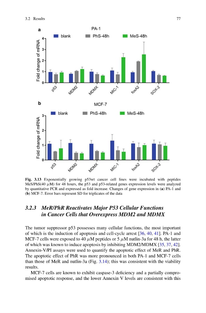
Fig. 3.13 Exponentially growing p53wt cancer cell lines were incubated with peptides
MeS/PhS(40 μM) for 48 hours, the p53 and p53-related genes expression levels were analyzed
by quantitative PCR and expressed as fold increase. Changes of gene expression in (a) PA-1 and
(b) MCF-7. Error bars represent SD for triplicates of the data
3.2.3 MeR/PhR Reactivates Major P53 Cellular Functions
in Cancer Cells that Overexpress MDM2 and MDMX
The tumor suppressor p53 possesses many cellular functions, the most important
of which is the induction of apoptosis and cell-cycle arrest [36, 40, 41]. PA-1 and
MCF-7 cells were exposed to 40 µM peptides or 5 µM nutlin-3a for 48 h, the latter
of which was known to induce apoptosis by inhibiting MDM2/MDMX [35, 37, 42].
Annexin-V/PI assays were used to quantify the apoptotic effect of MeR and PhR.
The apoptotic effect of PhR was more pronounced in both PA-1 and MCF-7 cells
than those of MeR and nutlin-3a (Fig. 3.14); this was consistent with the viability
results.
MCF-7 cells are known to exhibit caspase-3 deficiency and a partially compromised apoptotic response, and the lower Annexin V levels are consistent with this
77
Fig. 3.13 Exponentially growing p53wt cancer cell lines were incubated with peptides
MeS/PhS(40 μM) for 48 hours, the p53 and p53-related genes expression levels were analyzed
by quantitative PCR and expressed as fold increase. Changes of gene expression in (a) PA-1 and
(b) MCF-7. Error bars represent SD for triplicates of the data
3.2.3 MeR/PhR Reactivates Major P53 Cellular Functions
in Cancer Cells that Overexpress MDM2 and MDMX
The tumor suppressor p53 possesses many cellular functions, the most important
of which is the induction of apoptosis and cell-cycle arrest [36, 40, 41]. PA-1 and
MCF-7 cells were exposed to 40 µM peptides or 5 µM nutlin-3a for 48 h, the latter
of which was known to induce apoptosis by inhibiting MDM2/MDMX [35, 37, 42].
Annexin-V/PI assays were used to quantify the apoptotic effect of MeR and PhR.
The apoptotic effect of PhR was more pronounced in both PA-1 and MCF-7 cells
than those of MeR and nutlin-3a (Fig. 3.14); this was consistent with the viability
results.
MCF-7 cells are known to exhibit caspase-3 deficiency and a partially compromised apoptotic response, and the lower Annexin V levels are consistent with this