

3.2 Results
75
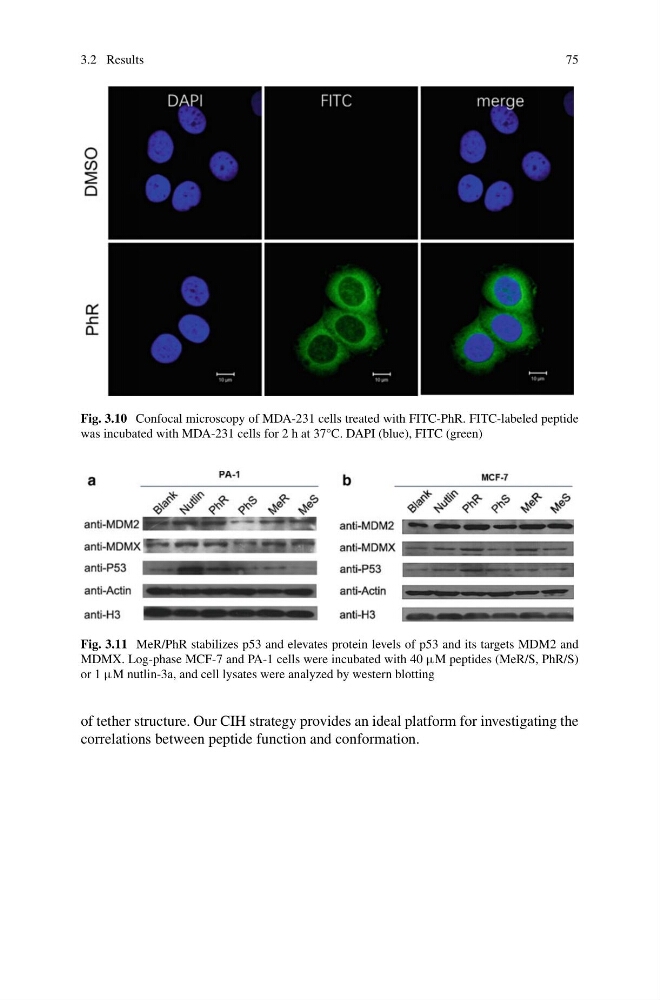
Fig. 3.10 Confocal microscopy of MDA-231 cells treated with FITC-PhR. FITC-labeled peptide
was incubated with MDA-231 cells for 2 h at 37°C. DAPI (blue), FITC (green)
Fig. 3.11 MeR/PhR stabilizes p53 and elevates protein levels of p53 and its targets MDM2 and
MDMX. Log-phase MCF-7 and PA-1 cells were incubated with 40 μM peptides (MeR/S, PhR/S)
or 1 μM nutlin-3a, and cell lysates were analyzed by western blotting
of tether structure. Our CIH strategy provides an ideal platform for investigating the
correlations between peptide function and conformation.
75
Fig. 3.10 Confocal microscopy of MDA-231 cells treated with FITC-PhR. FITC-labeled peptide
was incubated with MDA-231 cells for 2 h at 37°C. DAPI (blue), FITC (green)
Fig. 3.11 MeR/PhR stabilizes p53 and elevates protein levels of p53 and its targets MDM2 and
MDMX. Log-phase MCF-7 and PA-1 cells were incubated with 40 μM peptides (MeR/S, PhR/S)
or 1 μM nutlin-3a, and cell lysates were analyzed by western blotting
of tether structure. Our CIH strategy provides an ideal platform for investigating the
correlations between peptide function and conformation.