

72
3 In-Tether Chiral Center Induced Helical Peptide Modulators …
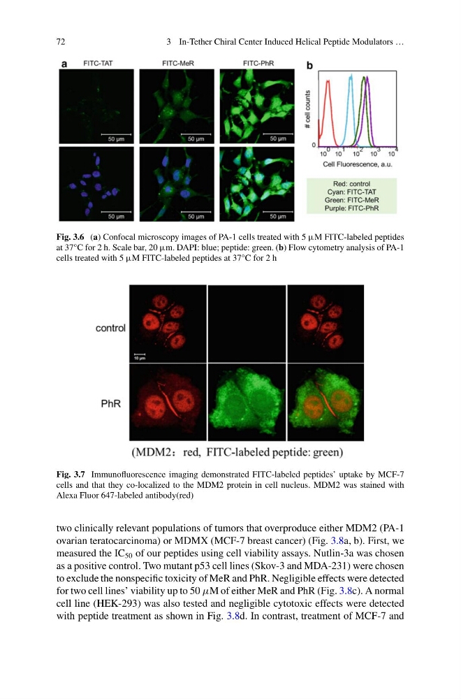
Fig. 3.6 (a) Confocal microscopy images of PA-1 cells treated with 5 μM FITC-labeled peptides
at 37°C for 2 h. Scale bar, 20 μm. DAPI: blue; peptide: green. (b) Flow cytometry analysis of PA-1
cells treated with 5 μM FITC-labeled peptides at 37°C for 2 h
Fig. 3.7 Immunofluorescence imaging demonstrated FITC-labeled peptides’ uptake by MCF-7
cells and that they co-localized to the MDM2 protein in cell nucleus. MDM2 was stained with
Alexa Fluor 647-labeled antibody(red)
two clinically relevant populations of tumors that overproduce either MDM2 (PA-1
ovarian teratocarcinoma) or MDMX (MCF-7 breast cancer) (Fig. 3.8a, b). First, we
measured the IC 50 of our peptides using cell viability assays. Nutlin-3a was chosen
as a positive control. Two mutant p53 cell lines (Skov-3 and MDA-231) were chosen
to exclude the nonspecific toxicity of MeR and PhR. Negligible effects were detected
for two cell lines’ viability up to 50 µM of either MeR and PhR (Fig. 3.8c). A normal
cell line (HEK-293) was also tested and negligible cytotoxic effects were detected
with peptide treatment as shown in Fig. 3.8d. In contrast, treatment of MCF-7 and
3 In-Tether Chiral Center Induced Helical Peptide Modulators …
Fig. 3.6 (a) Confocal microscopy images of PA-1 cells treated with 5 μM FITC-labeled peptides
at 37°C for 2 h. Scale bar, 20 μm. DAPI: blue; peptide: green. (b) Flow cytometry analysis of PA-1
cells treated with 5 μM FITC-labeled peptides at 37°C for 2 h
Fig. 3.7 Immunofluorescence imaging demonstrated FITC-labeled peptides’ uptake by MCF-7
cells and that they co-localized to the MDM2 protein in cell nucleus. MDM2 was stained with
Alexa Fluor 647-labeled antibody(red)
two clinically relevant populations of tumors that overproduce either MDM2 (PA-1
ovarian teratocarcinoma) or MDMX (MCF-7 breast cancer) (Fig. 3.8a, b). First, we
measured the IC 50 of our peptides using cell viability assays. Nutlin-3a was chosen
as a positive control. Two mutant p53 cell lines (Skov-3 and MDA-231) were chosen
to exclude the nonspecific toxicity of MeR and PhR. Negligible effects were detected
for two cell lines’ viability up to 50 µM of either MeR and PhR (Fig. 3.8c). A normal
cell line (HEK-293) was also tested and negligible cytotoxic effects were detected
with peptide treatment as shown in Fig. 3.8d. In contrast, treatment of MCF-7 and