

3.2 Results
71
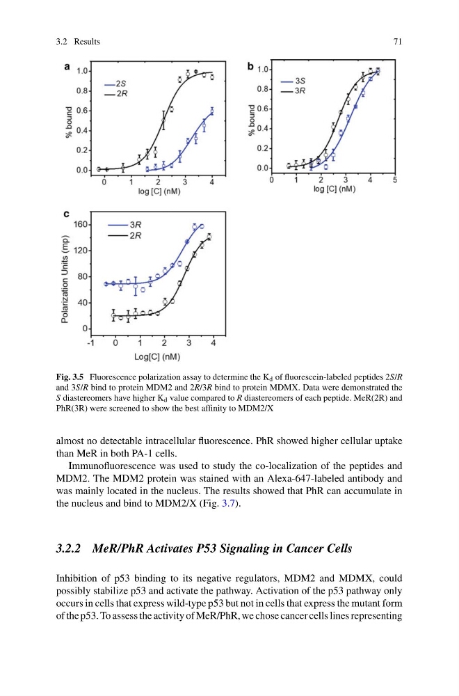
Fig. 3.5 Fluorescence polarization assay to determine the K d of fluorescein-labeled peptides 2S/R
and 3S/R bind to protein MDM2 and 2R/3R bind to protein MDMX. Data were demonstrated the
S diastereomers have higher K d value compared to R diastereomers of each peptide. MeR(2R) and
PhR(3R) were screened to show the best affinity to MDM2/X
almost no detectable intracellular fluorescence. PhR showed higher cellular uptake
than MeR in both PA-1 cells.
Immunofluorescence was used to study the co-localization of the peptides and
MDM2. The MDM2 protein was stained with an Alexa-647-labeled antibody and
was mainly located in the nucleus. The results showed that PhR can accumulate in
the nucleus and bind to MDM2/X (Fig. 3.7).
3.2.2 MeR/PhR Activates P53 Signaling in Cancer Cells
Inhibition of p53 binding to its negative regulators, MDM2 and MDMX, could
possibly stabilize p53 and activate the pathway. Activation of the p53 pathway only
occurs in cells that express wild-type p53 but not in cells that express the mutant form
of the p53. To assess the activity of MeR/PhR, we chose cancer cells lines representing
71
Fig. 3.5 Fluorescence polarization assay to determine the K d of fluorescein-labeled peptides 2S/R
and 3S/R bind to protein MDM2 and 2R/3R bind to protein MDMX. Data were demonstrated the
S diastereomers have higher K d value compared to R diastereomers of each peptide. MeR(2R) and
PhR(3R) were screened to show the best affinity to MDM2/X
almost no detectable intracellular fluorescence. PhR showed higher cellular uptake
than MeR in both PA-1 cells.
Immunofluorescence was used to study the co-localization of the peptides and
MDM2. The MDM2 protein was stained with an Alexa-647-labeled antibody and
was mainly located in the nucleus. The results showed that PhR can accumulate in
the nucleus and bind to MDM2/X (Fig. 3.7).
3.2.2 MeR/PhR Activates P53 Signaling in Cancer Cells
Inhibition of p53 binding to its negative regulators, MDM2 and MDMX, could
possibly stabilize p53 and activate the pathway. Activation of the p53 pathway only
occurs in cells that express wild-type p53 but not in cells that express the mutant form
of the p53. To assess the activity of MeR/PhR, we chose cancer cells lines representing