

2.2 Results
43
13a
13b
DAPI
Bright field
Merge
# Cells
FITC
FITC
20mm
a)
b)
c)
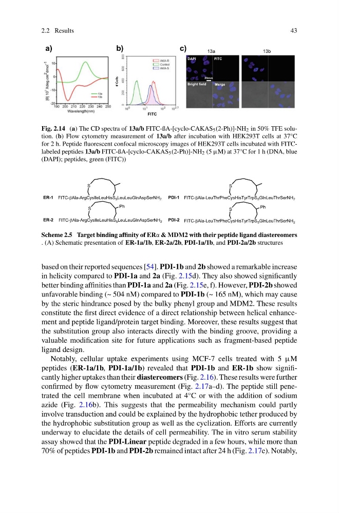
Fig. 2.14 (a) The CD spectra of 13a/b FITC-ßA-[cyclo-CAKAS 5 (2-Ph)]-NH 2 in 50% TFE solution. (b) Flow cytometry measurement of 13a/b after incubation with HEK293T cells at 37°C
for 2 h. Peptide fluorescent confocal microscopy images of HEK293T cells incubated with FITClabeled peptides 13a/b FITC-ßA-[cyclo-CAKAS 5 (2-Ph)]-NH 2 (5 μM) at 37°C for 1 h (DNA, blue
(DAPI); peptides, green (FITC))
Scheme 2.5 Target binding affinity of ERα & MDM2 with their peptide ligand diastereomers
. (A) Schematic presentation of ER-1a/1b, ER-2a/2b, PDI-1a/1b, and PDI-2a/2b structures
based on their reported sequences [54]. PDI-1b and 2b showed a remarkable increase
in helicity compared to PDI-1a and 2a (Fig. 2.15d). They also showed significantly
better binding affinities than PDI-1a and 2a (Fig. 2.15e, f). However, PDI-2b showed
unfavorable binding (~ 504 nM) compared to PDI-1b (~ 165 nM), which may cause
by the steric hindrance posed by the bulky phenyl group and MDM2. These results
constitute the first direct evidence of a direct relationship between helical enhancement and peptide ligand/protein target binding. Moreover, these results suggest that
the substitution group also interacts directly with the binding groove, providing a
valuable modification site for future applications such as fragment-based peptide
ligand design.
Notably, cellular uptake experiments using MCF-7 cells treated with 5 μM
peptides (ER-1a/1b, PDI-1a/1b) revealed that PDI-1b and ER-1b show significantly higher uptakes than their diastereomers (Fig. 2.16). These results were further
confirmed by flow cytometry measurement (Fig. 2.17a–d). The peptide still penetrated the cell membrane when incubated at 4°C or with the addition of sodium
azide (Fig. 2.16b). This suggests that the permeability mechanism could partly
involve transduction and could be explained by the hydrophobic tether produced by
the hydrophobic substitution group as well as the cyclization. Efforts are currently
underway to elucidate the details of cell permeability. The in vitro serum stability
assay showed that the PDI-Linear peptide degraded in a few hours, while more than
70% of peptides PDI-1b and PDI-2b remained intact after 24 h (Fig. 2.17e). Notably,
43
13a
13b
DAPI
Bright field
Merge
# Cells
FITC
FITC
20mm
a)
b)
c)
Fig. 2.14 (a) The CD spectra of 13a/b FITC-ßA-[cyclo-CAKAS 5 (2-Ph)]-NH 2 in 50% TFE solution. (b) Flow cytometry measurement of 13a/b after incubation with HEK293T cells at 37°C
for 2 h. Peptide fluorescent confocal microscopy images of HEK293T cells incubated with FITClabeled peptides 13a/b FITC-ßA-[cyclo-CAKAS 5 (2-Ph)]-NH 2 (5 μM) at 37°C for 1 h (DNA, blue
(DAPI); peptides, green (FITC))
Scheme 2.5 Target binding affinity of ERα & MDM2 with their peptide ligand diastereomers
. (A) Schematic presentation of ER-1a/1b, ER-2a/2b, PDI-1a/1b, and PDI-2a/2b structures
based on their reported sequences [54]. PDI-1b and 2b showed a remarkable increase
in helicity compared to PDI-1a and 2a (Fig. 2.15d). They also showed significantly
better binding affinities than PDI-1a and 2a (Fig. 2.15e, f). However, PDI-2b showed
unfavorable binding (~ 504 nM) compared to PDI-1b (~ 165 nM), which may cause
by the steric hindrance posed by the bulky phenyl group and MDM2. These results
constitute the first direct evidence of a direct relationship between helical enhancement and peptide ligand/protein target binding. Moreover, these results suggest that
the substitution group also interacts directly with the binding groove, providing a
valuable modification site for future applications such as fragment-based peptide
ligand design.
Notably, cellular uptake experiments using MCF-7 cells treated with 5 μM
peptides (ER-1a/1b, PDI-1a/1b) revealed that PDI-1b and ER-1b show significantly higher uptakes than their diastereomers (Fig. 2.16). These results were further
confirmed by flow cytometry measurement (Fig. 2.17a–d). The peptide still penetrated the cell membrane when incubated at 4°C or with the addition of sodium
azide (Fig. 2.16b). This suggests that the permeability mechanism could partly
involve transduction and could be explained by the hydrophobic tether produced by
the hydrophobic substitution group as well as the cyclization. Efforts are currently
underway to elucidate the details of cell permeability. The in vitro serum stability
assay showed that the PDI-Linear peptide degraded in a few hours, while more than
70% of peptides PDI-1b and PDI-2b remained intact after 24 h (Fig. 2.17e). Notably,