

42
2 Synthesis of In-Tether Chiral Center Peptides …
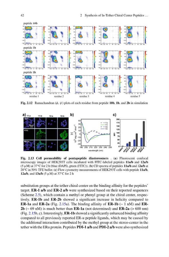
Fig. 2.12 Ramachandran (φ, ψ) plots of each residue from peptide 10b, 1b, and 2b in simulation
b)
c)
a)
12-a
12-b
11-a
11-b
DAPI
FITC
Merge
20um
Fig. 2.13 Cell permeability of pentapeptide diastereomers . (a) Fluorescent confocal
microscopy images of HEK293T cells incubated with FITC-labeled peptides 11a/b and 12a/b
(5 μM) at 37°C for 2 h (blue (DAPI), green (FITC)). (b) CD spectra of peptides 11a/b and 12a/b at
20°C in 50% TFE buffer. (c) Flow cytometry measurements of HEK293T cells with peptide 11a/b,
12a/b, and 13a/b (5 μM) at 37°C for 2 h
substitution groups at the tether chiral center on the binding affinity for the peptides’
target. ER-1 a/b and ER-2 a/b were synthesized based on their reported sequences
(Scheme 2.5), which contain a methyl or phenyl group at the chiral center, respectively. ER-1b and ER-2b showed a significant increase in helicity compared to
ER-1a and ER-2a (Fig. 2.15a). The binding affinity of ER-1b (~ 1 nM) and ER2b (~ 69 nM) is much better than ER-1a (not determined) and ER-2a (> 600 nm)
(Fig. 2.15b, c). Interestingly, ER-1b showed a significantly enhanced binding affinity
compared to all previously reported ER-α peptide ligands, which may be caused by
the additional interaction contributed by the methyl group at the stereo-center in the
tether with the ERα protein. Peptides PDI-1 a/b and PDI-2 a/b were also synthesized
2 Synthesis of In-Tether Chiral Center Peptides …
Fig. 2.12 Ramachandran (φ, ψ) plots of each residue from peptide 10b, 1b, and 2b in simulation
b)
c)
a)
12-a
12-b
11-a
11-b
DAPI
FITC
Merge
20um
Fig. 2.13 Cell permeability of pentapeptide diastereomers . (a) Fluorescent confocal
microscopy images of HEK293T cells incubated with FITC-labeled peptides 11a/b and 12a/b
(5 μM) at 37°C for 2 h (blue (DAPI), green (FITC)). (b) CD spectra of peptides 11a/b and 12a/b at
20°C in 50% TFE buffer. (c) Flow cytometry measurements of HEK293T cells with peptide 11a/b,
12a/b, and 13a/b (5 μM) at 37°C for 2 h
substitution groups at the tether chiral center on the binding affinity for the peptides’
target. ER-1 a/b and ER-2 a/b were synthesized based on their reported sequences
(Scheme 2.5), which contain a methyl or phenyl group at the chiral center, respectively. ER-1b and ER-2b showed a significant increase in helicity compared to
ER-1a and ER-2a (Fig. 2.15a). The binding affinity of ER-1b (~ 1 nM) and ER2b (~ 69 nM) is much better than ER-1a (not determined) and ER-2a (> 600 nm)
(Fig. 2.15b, c). Interestingly, ER-1b showed a significantly enhanced binding affinity
compared to all previously reported ER-α peptide ligands, which may be caused by
the additional interaction contributed by the methyl group at the stereo-center in the
tether with the ERα protein. Peptides PDI-1 a/b and PDI-2 a/b were also synthesized