

2.2 Results
33
a
365nm h.v.
2h, DMF
Ac--C--X--X--X--S 5 -
SH
R
COOH
NH 2
Me
S 5 (2-Me):
Cyclo-Ac-CAAAS 5 (2-Me)-NH 2
1a and 1b
b
Ac--C--X--X--X--S 5 -
S
R
c
d
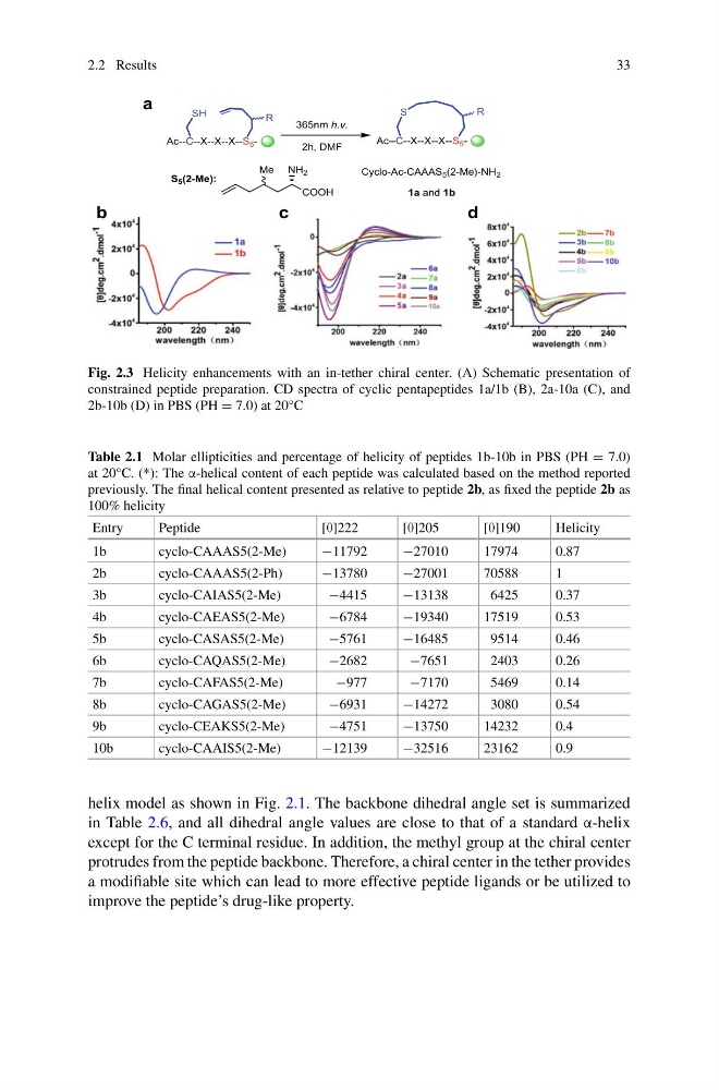
Fig. 2.3 Helicity enhancements with an in-tether chiral center. (A) Schematic presentation of
constrained peptide preparation. CD spectra of cyclic pentapeptides 1a/1b (B), 2a-10a (C), and
2b-10b (D) in PBS (PH = 7.0) at 20°C
Table 2.1 Molar ellipticities and percentage of helicity of peptides 1b-10b in PBS (PH = 7.0)
at 20°C. (*): The α-helical content of each peptide was calculated based on the method reported
previously. The final helical content presented as relative to peptide 2b, as fixed the peptide 2b as
100% helicity
Entry
Peptide
[θ]222
[θ]205
[θ]190
Helicity
1b
cyclo-CAAAS5(2-Me)
−11792
−27010
17974
0.87
2b
cyclo-CAAAS5(2-Ph)
−13780
−27001
70588
1
3b
cyclo-CAIAS5(2-Me)
−4415
−13138
6425
0.37
4b
cyclo-CAEAS5(2-Me)
−6784
−19340
17519
0.53
5b
cyclo-CASAS5(2-Me)
−5761
−16485
9514
0.46
6b
cyclo-CAQAS5(2-Me)
−2682
−7651
2403
0.26
7b
cyclo-CAFAS5(2-Me)
−977
−7170
5469
0.14
8b
cyclo-CAGAS5(2-Me)
−6931
−14272
3080
0.54
9b
cyclo-CEAKS5(2-Me)
−4751
−13750
14232
0.4
10b
cyclo-CAAIS5(2-Me)
−12139
−32516
23162
0.9
helix model as shown in Fig. 2.1. The backbone dihedral angle set is summarized
in Table 2.6, and all dihedral angle values are close to that of a standard α-helix
except for the C terminal residue. In addition, the methyl group at the chiral center
protrudes from the peptide backbone. Therefore, a chiral center in the tether provides
a modifiable site which can lead to more effective peptide ligands or be utilized to
improve the peptide’s drug-like property.
33
a
365nm h.v.
2h, DMF
Ac--C--X--X--X--S 5 -
SH
R
COOH
NH 2
Me
S 5 (2-Me):
Cyclo-Ac-CAAAS 5 (2-Me)-NH 2
1a and 1b
b
Ac--C--X--X--X--S 5 -
S
R
c
d
Fig. 2.3 Helicity enhancements with an in-tether chiral center. (A) Schematic presentation of
constrained peptide preparation. CD spectra of cyclic pentapeptides 1a/1b (B), 2a-10a (C), and
2b-10b (D) in PBS (PH = 7.0) at 20°C
Table 2.1 Molar ellipticities and percentage of helicity of peptides 1b-10b in PBS (PH = 7.0)
at 20°C. (*): The α-helical content of each peptide was calculated based on the method reported
previously. The final helical content presented as relative to peptide 2b, as fixed the peptide 2b as
100% helicity
Entry
Peptide
[θ]222
[θ]205
[θ]190
Helicity
1b
cyclo-CAAAS5(2-Me)
−11792
−27010
17974
0.87
2b
cyclo-CAAAS5(2-Ph)
−13780
−27001
70588
1
3b
cyclo-CAIAS5(2-Me)
−4415
−13138
6425
0.37
4b
cyclo-CAEAS5(2-Me)
−6784
−19340
17519
0.53
5b
cyclo-CASAS5(2-Me)
−5761
−16485
9514
0.46
6b
cyclo-CAQAS5(2-Me)
−2682
−7651
2403
0.26
7b
cyclo-CAFAS5(2-Me)
−977
−7170
5469
0.14
8b
cyclo-CAGAS5(2-Me)
−6931
−14272
3080
0.54
9b
cyclo-CEAKS5(2-Me)
−4751
−13750
14232
0.4
10b
cyclo-CAAIS5(2-Me)
−12139
−32516
23162
0.9
helix model as shown in Fig. 2.1. The backbone dihedral angle set is summarized
in Table 2.6, and all dihedral angle values are close to that of a standard α-helix
except for the C terminal residue. In addition, the methyl group at the chiral center
protrudes from the peptide backbone. Therefore, a chiral center in the tether provides
a modifiable site which can lead to more effective peptide ligands or be utilized to
improve the peptide’s drug-like property.