

32
2 Synthesis of In-Tether Chiral Center Peptides …
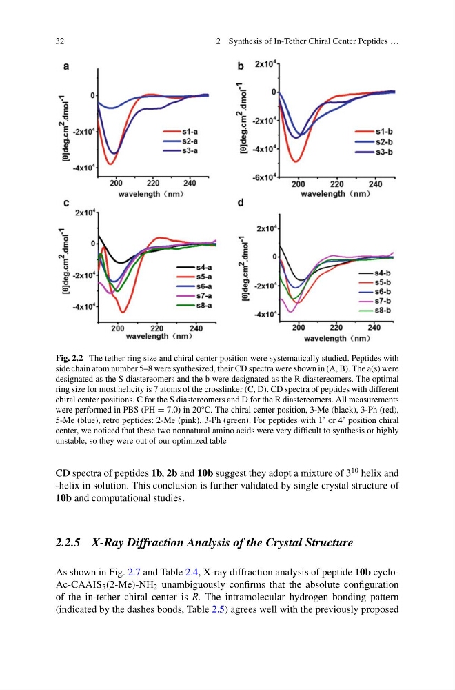
Fig. 2.2 The tether ring size and chiral center position were systematically studied. Peptides with
side chain atom number 5–8 were synthesized, their CD spectra were shown in (A, B). The a(s) were
designated as the S diastereomers and the b were designated as the R diastereomers. The optimal
ring size for most helicity is 7 atoms of the crosslinker (C, D). CD spectra of peptides with different
chiral center positions. C for the S diastereomers and D for the R diastereomers. All measurements
were performed in PBS (PH = 7.0) in 20°C. The chiral center position, 3-Me (black), 3-Ph (red),
5-Me (blue), retro peptides: 2-Me (pink), 3-Ph (green). For peptides with 1’ or 4’ position chiral
center, we noticed that these two nonnatural amino acids were very difficult to synthesis or highly
unstable, so they were out of our optimized table
CD spectra of peptides 1b, 2b and 10b suggest they adopt a mixture of 3
10 helix and
-helix in solution. This conclusion is further validated by single crystal structure of
10b and computational studies.
2.2.5 X-Ray Diffraction Analysis of the Crystal Structure
As shown in Fig. 2.7 and Table 2.4, X-ray diffraction analysis of peptide 10b cycloAc-CAAIS 5 (2-Me)-NH 2 unambiguously confirms that the absolute configuration
of the in-tether chiral center is R. The intramolecular hydrogen bonding pattern
(indicated by the dashes bonds, Table 2.5) agrees well with the previously proposed
2 Synthesis of In-Tether Chiral Center Peptides …
Fig. 2.2 The tether ring size and chiral center position were systematically studied. Peptides with
side chain atom number 5–8 were synthesized, their CD spectra were shown in (A, B). The a(s) were
designated as the S diastereomers and the b were designated as the R diastereomers. The optimal
ring size for most helicity is 7 atoms of the crosslinker (C, D). CD spectra of peptides with different
chiral center positions. C for the S diastereomers and D for the R diastereomers. All measurements
were performed in PBS (PH = 7.0) in 20°C. The chiral center position, 3-Me (black), 3-Ph (red),
5-Me (blue), retro peptides: 2-Me (pink), 3-Ph (green). For peptides with 1’ or 4’ position chiral
center, we noticed that these two nonnatural amino acids were very difficult to synthesis or highly
unstable, so they were out of our optimized table
CD spectra of peptides 1b, 2b and 10b suggest they adopt a mixture of 3
10 helix and
-helix in solution. This conclusion is further validated by single crystal structure of
10b and computational studies.
2.2.5 X-Ray Diffraction Analysis of the Crystal Structure
As shown in Fig. 2.7 and Table 2.4, X-ray diffraction analysis of peptide 10b cycloAc-CAAIS 5 (2-Me)-NH 2 unambiguously confirms that the absolute configuration
of the in-tether chiral center is R. The intramolecular hydrogen bonding pattern
(indicated by the dashes bonds, Table 2.5) agrees well with the previously proposed