

2.2 Results
31
Ac-C--A--A--A--S 5 -NH 2
S
Me
Ac-S 5 --A--A--A--C-NH 2
Ac-S 5 --A--A--A--C-NH 2
S
Me
Ac-C--A--A--A--S 5 -NH 2
S
Me
Ac-C--A--A--A--S 5 -NH 2
S
Me
Ac-C--A--A--A--S 5 -NH 2
S
S4-a/b
S6-a/b
S8-a/b
S1-a/b
S2-a/b
S3-a/b
Me
Ac-C--A--A--A--S 5 -NH 2
S5-a/b
Ring size:
Chiral centre position:
Retro peptide:
Ac-C--A--A--A--S 5 -NH 2
S7-a/b
S
Ph
S
Me
S
Ph
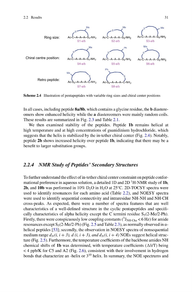
Scheme 2.4 Illustration of pentapeptides with variable ring sizes and chiral center positions
In all cases, including peptide 8a/8b, which contains a glycine residue, the b diastereomers show enhanced helicity while the a diastereomers were mainly random coils.
These results are summarized in Fig. 2.3 and Table 2.1.
We then examined stability of the peptides. Peptide 1b remains helical at
high temperature and at high concentrations of guanidinium hydrochloride, which
suggests that the helix is stabilized by the in-tether chiral center (Fig. 2.4). Notably,
peptide 2b shows increased helicity over peptide 1b, indicating that there may be a
benefit to larger substitution groups.
2.2.4 NMR Study of Peptides’ Secondary Structures
To further understand the effect of in-tether chiral center constraint on peptide conformational preference in aqueous solution, a detailed 1D and 2D
1 H-NMR study of 1b,
2b, and 10b was performed in 10% D 2 O in H 2 O at 25°C. 2D-TOCSY spectra were
used to identify resonances for each amino acid (Table 2.2), and NOESY spectra
were used to identify sequential connectivity and intraresidue NH-NH and NH-CH
cross-peaks. As expected, there were a number of spectra features that are well
characteristics of a well-defined structure in the cyclic pentapeptides and specifically characteristics of alpha helicity except the C termini residue S 5 (2-Me/2-Ph).
Firstly, there were conspicuously low coupling constants (
3 J NH-Cha < 6 Hz) for amide
resonances except S 5 (2-Me/2-Ph) (Fig. 2.5 and Table 2.3), as normally observed in αhelical peptides [53]; secondly, the observation in NOESY spectra of nonsequential
medium range d N (i, i + 3), d (i, i + 3), and d N (i, i + 4) NOEs suggest helical structure (Fig. 2.5). Furthermore, the temperature coefficients of the backbone amides NH
chemical shifts of 1b was determined, with temperature coefficients ( being
< 4 ppb/K for C5 and A2 (Fig. 2.6), consistent with their involvement in hydrogen
bonds that characterize an -helix or 3
10 helix. In summary, the NOE spectrums and
31
Ac-C--A--A--A--S 5 -NH 2
S
Me
Ac-S 5 --A--A--A--C-NH 2
Ac-S 5 --A--A--A--C-NH 2
S
Me
Ac-C--A--A--A--S 5 -NH 2
S
Me
Ac-C--A--A--A--S 5 -NH 2
S
Me
Ac-C--A--A--A--S 5 -NH 2
S
S4-a/b
S6-a/b
S8-a/b
S1-a/b
S2-a/b
S3-a/b
Me
Ac-C--A--A--A--S 5 -NH 2
S5-a/b
Ring size:
Chiral centre position:
Retro peptide:
Ac-C--A--A--A--S 5 -NH 2
S7-a/b
S
Ph
S
Me
S
Ph
Scheme 2.4 Illustration of pentapeptides with variable ring sizes and chiral center positions
In all cases, including peptide 8a/8b, which contains a glycine residue, the b diastereomers show enhanced helicity while the a diastereomers were mainly random coils.
These results are summarized in Fig. 2.3 and Table 2.1.
We then examined stability of the peptides. Peptide 1b remains helical at
high temperature and at high concentrations of guanidinium hydrochloride, which
suggests that the helix is stabilized by the in-tether chiral center (Fig. 2.4). Notably,
peptide 2b shows increased helicity over peptide 1b, indicating that there may be a
benefit to larger substitution groups.
2.2.4 NMR Study of Peptides’ Secondary Structures
To further understand the effect of in-tether chiral center constraint on peptide conformational preference in aqueous solution, a detailed 1D and 2D
1 H-NMR study of 1b,
2b, and 10b was performed in 10% D 2 O in H 2 O at 25°C. 2D-TOCSY spectra were
used to identify resonances for each amino acid (Table 2.2), and NOESY spectra
were used to identify sequential connectivity and intraresidue NH-NH and NH-CH
cross-peaks. As expected, there were a number of spectra features that are well
characteristics of a well-defined structure in the cyclic pentapeptides and specifically characteristics of alpha helicity except the C termini residue S 5 (2-Me/2-Ph).
Firstly, there were conspicuously low coupling constants (
3 J NH-Cha < 6 Hz) for amide
resonances except S 5 (2-Me/2-Ph) (Fig. 2.5 and Table 2.3), as normally observed in αhelical peptides [53]; secondly, the observation in NOESY spectra of nonsequential
medium range d N (i, i + 3), d (i, i + 3), and d N (i, i + 4) NOEs suggest helical structure (Fig. 2.5). Furthermore, the temperature coefficients of the backbone amides NH
chemical shifts of 1b was determined, with temperature coefficients ( being
< 4 ppb/K for C5 and A2 (Fig. 2.6), consistent with their involvement in hydrogen
bonds that characterize an -helix or 3
10 helix. In summary, the NOE spectrums and