

1.1 Introduction of Protein-Protein Interactions
3
N
N
N
N
N
N
O
O
O
O
O
O
H
H
H
H
H
H
i
i + 1
i+2
i+3
i+4
i+5
helix H-bond patterns:
3 10
-helix
3.6 13
-helix
4.4 16
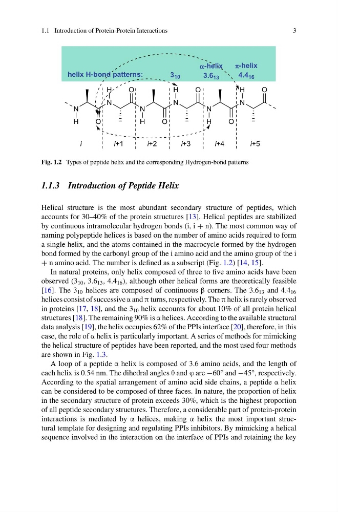
Fig. 1.2 Types of peptide helix and the corresponding Hydrogen-bond patterns
1.1.3 Introduction of Peptide Helix
Helical structure is the most abundant secondary structure of peptides, which
accounts for 30–40% of the protein structures [13]. Helical peptides are stabilized
by continuous intramolecular hydrogen bonds (i, i + n). The most common way of
naming polypeptide helices is based on the number of amino acids required to form
a single helix, and the atoms contained in the macrocycle formed by the hydrogen
bond formed by the carbonyl group of the i amino acid and the amino group of the i
+ n amino acid. The number is defined as a subscript (Fig. 1.2) [14, 15].
In natural proteins, only helix composed of three to five amino acids have been
observed (3 10 , 3.6 13 , 4.4 16 ), although other helical forms are theoretically feasible
[16]. The 3 10 helices are composed of continuous β corners. The 3.6 13 and 4.4 16
helices consist of successive α and π turns, respectively. The π helix is rarely observed
in proteins [17, 18], and the 3 10 helix accounts for about 10% of all protein helical
structures [18]. The remaining 90% is α helices. According to the available structural
data analysis [19], the helix occupies 62% of the PPIs interface [20], therefore, in this
case, the role of α helix is particularly important. A series of methods for mimicking
the helical structure of peptides have been reported, and the most used four methods
are shown in Fig. 1.3.
A loop of a peptide α helix is composed of 3.6 amino acids, and the length of
each helix is 0.54 nm. The dihedral angles θ and ϕ are −60° and −45°, respectively.
According to the spatial arrangement of amino acid side chains, a peptide α helix
can be considered to be composed of three faces. In nature, the proportion of helix
in the secondary structure of protein exceeds 30%, which is the highest proportion
of all peptide secondary structures. Therefore, a considerable part of protein-protein
interactions is mediated by α helices, making α helix the most important structural template for designing and regulating PPIs inhibitors. By mimicking a helical
sequence involved in the interaction on the interface of PPIs and retaining the key
3
N
N
N
N
N
N
O
O
O
O
O
O
H
H
H
H
H
H
i
i + 1
i+2
i+3
i+4
i+5
helix H-bond patterns:
3 10
-helix
3.6 13
-helix
4.4 16
Fig. 1.2 Types of peptide helix and the corresponding Hydrogen-bond patterns
1.1.3 Introduction of Peptide Helix
Helical structure is the most abundant secondary structure of peptides, which
accounts for 30–40% of the protein structures [13]. Helical peptides are stabilized
by continuous intramolecular hydrogen bonds (i, i + n). The most common way of
naming polypeptide helices is based on the number of amino acids required to form
a single helix, and the atoms contained in the macrocycle formed by the hydrogen
bond formed by the carbonyl group of the i amino acid and the amino group of the i
+ n amino acid. The number is defined as a subscript (Fig. 1.2) [14, 15].
In natural proteins, only helix composed of three to five amino acids have been
observed (3 10 , 3.6 13 , 4.4 16 ), although other helical forms are theoretically feasible
[16]. The 3 10 helices are composed of continuous β corners. The 3.6 13 and 4.4 16
helices consist of successive α and π turns, respectively. The π helix is rarely observed
in proteins [17, 18], and the 3 10 helix accounts for about 10% of all protein helical
structures [18]. The remaining 90% is α helices. According to the available structural
data analysis [19], the helix occupies 62% of the PPIs interface [20], therefore, in this
case, the role of α helix is particularly important. A series of methods for mimicking
the helical structure of peptides have been reported, and the most used four methods
are shown in Fig. 1.3.
A loop of a peptide α helix is composed of 3.6 amino acids, and the length of
each helix is 0.54 nm. The dihedral angles θ and ϕ are −60° and −45°, respectively.
According to the spatial arrangement of amino acid side chains, a peptide α helix
can be considered to be composed of three faces. In nature, the proportion of helix
in the secondary structure of protein exceeds 30%, which is the highest proportion
of all peptide secondary structures. Therefore, a considerable part of protein-protein
interactions is mediated by α helices, making α helix the most important structural template for designing and regulating PPIs inhibitors. By mimicking a helical
sequence involved in the interaction on the interface of PPIs and retaining the key