

185
The low EC50-48 h values are coherent with findings in the literature and can be
categorized as “very highly toxic” to the test organisms. For example, Asghari et al.
(2012) found an EC50 = 2 μg L
−1
on D. magna for spherical silver nanoparticles of
16.6 nm.
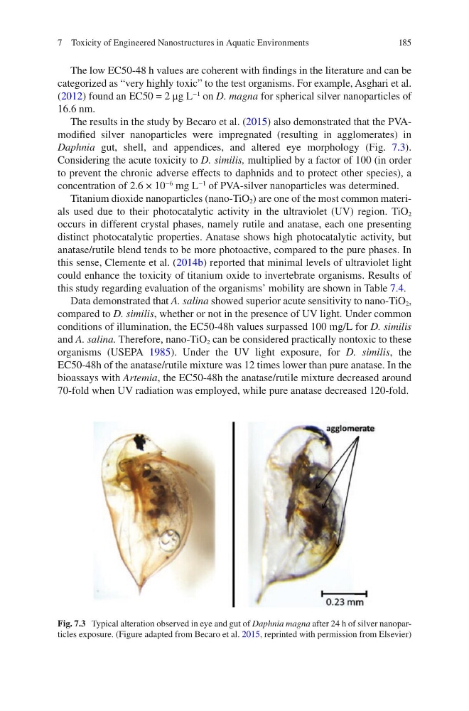
The results in the study by Becaro et al. (2015) also demonstrated that the PVAmodified silver nanoparticles were impregnated (resulting in agglomerates) in
Daphnia gut, shell, and appendices, and altered eye morphology (Fig. 7.3).
Considering the acute toxicity to D. similis, multiplied by a factor of 100 (in order
to prevent the chronic adverse effects to daphnids and to protect other species), a
concentration of 2.6 × 10
−6
mg L
−1
of PVA-silver nanoparticles was determined.
Titanium dioxide nanoparticles (nano-TiO 2 ) are one of the most common materials used due to their photocatalytic activity in the ultraviolet (UV) region. TiO 2
occurs in different crystal phases, namely rutile and anatase, each one presenting
distinct photocatalytic properties. Anatase shows high photocatalytic activity, but
anatase/rutile blend tends to be more photoactive, compared to the pure phases. In
this sense, Clemente et al. (2014b) reported that minimal levels of ultraviolet light
could enhance the toxicity of titanium oxide to invertebrate organisms. Results of
this study regarding evaluation of the organisms’ mobility are shown in Table 7.4.
Data demonstrated that A. salina showed superior acute sensitivity to nano-TiO 2 ,
compared to D. similis, whether or not in the presence of UV light. Under common
conditions of illumination, the EC50-48h values surpassed 100 mg/L for D. similis
and A. salina. Therefore, nano-TiO 2 can be considered practically nontoxic to these
organisms (USEPA 1985). Under the UV light exposure, for D. similis, the
EC50-48h of the anatase/rutile mixture was 12 times lower than pure anatase. In the
bioassays with Artemia, the EC50-48h the anatase/rutile mixture decreased around
70-fold when UV radiation was employed, while pure anatase decreased 120-fold.
Fig. 7.3 Typical alteration observed in eye and gut of Daphnia magna after 24 h of silver nanoparticles exposure. (Figure adapted from Becaro et al. 2015, reprinted with permission from Elsevier)
7 Toxicity of Engineered Nanostructures in Aquatic Environments
The low EC50-48 h values are coherent with findings in the literature and can be
categorized as “very highly toxic” to the test organisms. For example, Asghari et al.
(2012) found an EC50 = 2 μg L
−1
on D. magna for spherical silver nanoparticles of
16.6 nm.
The results in the study by Becaro et al. (2015) also demonstrated that the PVAmodified silver nanoparticles were impregnated (resulting in agglomerates) in
Daphnia gut, shell, and appendices, and altered eye morphology (Fig. 7.3).
Considering the acute toxicity to D. similis, multiplied by a factor of 100 (in order
to prevent the chronic adverse effects to daphnids and to protect other species), a
concentration of 2.6 × 10
−6
mg L
−1
of PVA-silver nanoparticles was determined.
Titanium dioxide nanoparticles (nano-TiO 2 ) are one of the most common materials used due to their photocatalytic activity in the ultraviolet (UV) region. TiO 2
occurs in different crystal phases, namely rutile and anatase, each one presenting
distinct photocatalytic properties. Anatase shows high photocatalytic activity, but
anatase/rutile blend tends to be more photoactive, compared to the pure phases. In
this sense, Clemente et al. (2014b) reported that minimal levels of ultraviolet light
could enhance the toxicity of titanium oxide to invertebrate organisms. Results of
this study regarding evaluation of the organisms’ mobility are shown in Table 7.4.
Data demonstrated that A. salina showed superior acute sensitivity to nano-TiO 2 ,
compared to D. similis, whether or not in the presence of UV light. Under common
conditions of illumination, the EC50-48h values surpassed 100 mg/L for D. similis
and A. salina. Therefore, nano-TiO 2 can be considered practically nontoxic to these
organisms (USEPA 1985). Under the UV light exposure, for D. similis, the
EC50-48h of the anatase/rutile mixture was 12 times lower than pure anatase. In the
bioassays with Artemia, the EC50-48h the anatase/rutile mixture decreased around
70-fold when UV radiation was employed, while pure anatase decreased 120-fold.
Fig. 7.3 Typical alteration observed in eye and gut of Daphnia magna after 24 h of silver nanoparticles exposure. (Figure adapted from Becaro et al. 2015, reprinted with permission from Elsevier)
7 Toxicity of Engineered Nanostructures in Aquatic Environments