

184
Toxicity Assays with Microcrustaceans Daphnia and Artemia
Toxicity tests employing both aquatic arthropoda (Daphnia and Artemia) are widely
used to evaluate the potential risk of environmental contamination by nanomaterials
(Chen et al. 2017; Zhu et al. 2017; Novak et al. 2018; Johnson et al. 2017). These
microcrustaceans represent the invertebrates at the base of the food chain, so toxic
effects at this trophic level can compromise the entire ecosystem (Dorchin and
Shanas 2013; Yang et al. 2018).
Daphnia sp is a bioindicator organism listed in the “Manual of tests for evaluation of ecotoxicity of chemical agents” (IBAMA 1988) and used in studies for the
registration of chemicals in regulatory Brazilian agencies, as well as by international protocols (Organisation for Economic Co-operation and Development. 2004)
(OECD 2004) in nanomaterial assessment (Oleszczuk et al. 2015; Gao et al. 2018).
Artemia salina is a phytoplanktonic arthropod capable to respond to varied
aquatic pollutants. Although it is a representative of marine ecosystems, Artemia
has been used together with freshwater organisms in hazard concentration predictions of nanomaterials and others compounds (Nogueira et al. 2015; Castro
et al. 2018).
Based on a body growth test for 96 hours, Castro et al. (2018) determined the
EC50-96h of graphene oxide for D. magna. Among ten organisms evaluated for different trophic levels, the daphnids showed the highest sensibility to the nanomaterial with EC50-96h values of 0.58 (0.35–2.07) and 0.42 (0.076–1.09) mg/L in the
absence and in the presence of humic acid, respectively.
Adam et al. (2015) indicate that the toxicity of copper oxide nanoparticles to
D. magna is caused by copper ions formed during dissolution of the nanoparticles
in the exposure medium. The majority of the particles formed large aggregates
while a small fraction dissolved. This may explain the much lower effects of copper
oxide nanoparticles (EC50 reproduction = 1.04 mg Cu/l) compared to copper salts
(EC50 reproduction = 0.022 mg Cu/l). Although the authors found much higher
metal concentration when exposed to the nanoparticles than when exposed to the
salt, the Cu did not internalize in the tissues or cells to cause additional toxic effects
after adsorbed on the carapace or ingested by the organisms.
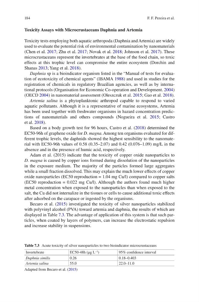
Becaro et al. (2015) investigated the toxicity of silver nanoparticles stabilized
with polyvinyl alcohol (PVA) toward artemia and daphnia, the results of which are
displayed in Table 7.3. The advantage of application of this system is that such particles, when coated by layers of polymers, can increase the electrostatic repulsion
and increase stability in suspensions.
Table 7.3 Acute toxicity of silver nanoparticles to two bioindicator microcrustaceans
Invertebrate
EC50-48h (μg L
−1 )
95% confidence interval
Daphnia similis
0.26
0.18–0.403
Artemia salina
55.0
22.0–11.0
Adapted from Becaro et al. (2015)
F. F. Pereira et al.
Toxicity Assays with Microcrustaceans Daphnia and Artemia
Toxicity tests employing both aquatic arthropoda (Daphnia and Artemia) are widely
used to evaluate the potential risk of environmental contamination by nanomaterials
(Chen et al. 2017; Zhu et al. 2017; Novak et al. 2018; Johnson et al. 2017). These
microcrustaceans represent the invertebrates at the base of the food chain, so toxic
effects at this trophic level can compromise the entire ecosystem (Dorchin and
Shanas 2013; Yang et al. 2018).
Daphnia sp is a bioindicator organism listed in the “Manual of tests for evaluation of ecotoxicity of chemical agents” (IBAMA 1988) and used in studies for the
registration of chemicals in regulatory Brazilian agencies, as well as by international protocols (Organisation for Economic Co-operation and Development. 2004)
(OECD 2004) in nanomaterial assessment (Oleszczuk et al. 2015; Gao et al. 2018).
Artemia salina is a phytoplanktonic arthropod capable to respond to varied
aquatic pollutants. Although it is a representative of marine ecosystems, Artemia
has been used together with freshwater organisms in hazard concentration predictions of nanomaterials and others compounds (Nogueira et al. 2015; Castro
et al. 2018).
Based on a body growth test for 96 hours, Castro et al. (2018) determined the
EC50-96h of graphene oxide for D. magna. Among ten organisms evaluated for different trophic levels, the daphnids showed the highest sensibility to the nanomaterial with EC50-96h values of 0.58 (0.35–2.07) and 0.42 (0.076–1.09) mg/L in the
absence and in the presence of humic acid, respectively.
Adam et al. (2015) indicate that the toxicity of copper oxide nanoparticles to
D. magna is caused by copper ions formed during dissolution of the nanoparticles
in the exposure medium. The majority of the particles formed large aggregates
while a small fraction dissolved. This may explain the much lower effects of copper
oxide nanoparticles (EC50 reproduction = 1.04 mg Cu/l) compared to copper salts
(EC50 reproduction = 0.022 mg Cu/l). Although the authors found much higher
metal concentration when exposed to the nanoparticles than when exposed to the
salt, the Cu did not internalize in the tissues or cells to cause additional toxic effects
after adsorbed on the carapace or ingested by the organisms.
Becaro et al. (2015) investigated the toxicity of silver nanoparticles stabilized
with polyvinyl alcohol (PVA) toward artemia and daphnia, the results of which are
displayed in Table 7.3. The advantage of application of this system is that such particles, when coated by layers of polymers, can increase the electrostatic repulsion
and increase stability in suspensions.
Table 7.3 Acute toxicity of silver nanoparticles to two bioindicator microcrustaceans
Invertebrate
EC50-48h (μg L
−1 )
95% confidence interval
Daphnia similis
0.26
0.18–0.403
Artemia salina
55.0
22.0–11.0
Adapted from Becaro et al. (2015)
F. F. Pereira et al.