

286
Z. Louna et al.
difference between osteons and the entire bone. Despite the development of many
continuum models of trabecular bone over the last two decades under the umbrella
of classical elasticity (e.g., Taylor et al. 2002; Bowman et al. 1998), those models
ignore microstructure-related scale effects on the macroscopic mechanical properties. They accordingly do not provide a satisfactory description of the bone behavior
when the microstructural size of bone is comparable to the macroscopic length scale.
It is accordingly the aim of the developed strain gradient continuum theory of bone to
remedy these deficiencies and to properly account for microstructural effects when
modeling the evolutive bone microstructure.
Strain gradient models for bone remodeling rely on micromechanical analyses
performed at the scale of a representative volume element of trabecular bone structure (Louna et al. 2018). Both static and evolutive homogenized properties of a
periodic network of bone trabeculae are evaluated by combining a methodology for
the evaluation of the average kinematic and static variables over a trabecular cell and
numerical simulations with controlled imposed first and second strain rates. In fact,
the use of a strain gradient model accounts explicitly for the precise microstructure of
the porous trabecular network, i.e., for the geometric distribution of porosity and the
size of the pores inside the representative volume element. The strain gradient type
constitutive model has been identified within the umbrella of the thermodynamics of
irreversible processes, adopting a split of the kinematic and static tensors into their
deviator and hydrostatic contributions. The elaborated effective growth constitutive
law at the scale of the homogenized set of trabeculae consequently relates the average
first and second growth strain rates to the homogenized Cauchy stress and hyperstress
tensors, weighted by a nonlinear function of the (evolving) apparent density.
We herewith summarize the constitutive model developed in (Louna et al. 2019).
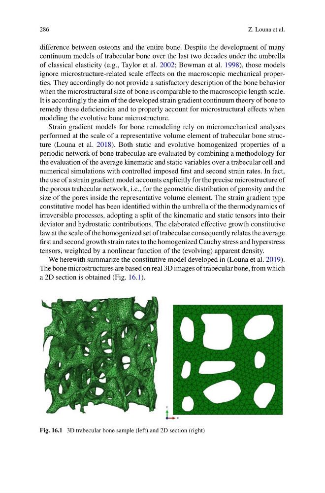
The bone microstructures are based on real 3D images of trabecular bone, from which
a 2D section is obtained (Fig. 16.1).
Fig. 16.1 3D trabecular bone sample (left) and 2D section (right)
Z. Louna et al.
difference between osteons and the entire bone. Despite the development of many
continuum models of trabecular bone over the last two decades under the umbrella
of classical elasticity (e.g., Taylor et al. 2002; Bowman et al. 1998), those models
ignore microstructure-related scale effects on the macroscopic mechanical properties. They accordingly do not provide a satisfactory description of the bone behavior
when the microstructural size of bone is comparable to the macroscopic length scale.
It is accordingly the aim of the developed strain gradient continuum theory of bone to
remedy these deficiencies and to properly account for microstructural effects when
modeling the evolutive bone microstructure.
Strain gradient models for bone remodeling rely on micromechanical analyses
performed at the scale of a representative volume element of trabecular bone structure (Louna et al. 2018). Both static and evolutive homogenized properties of a
periodic network of bone trabeculae are evaluated by combining a methodology for
the evaluation of the average kinematic and static variables over a trabecular cell and
numerical simulations with controlled imposed first and second strain rates. In fact,
the use of a strain gradient model accounts explicitly for the precise microstructure of
the porous trabecular network, i.e., for the geometric distribution of porosity and the
size of the pores inside the representative volume element. The strain gradient type
constitutive model has been identified within the umbrella of the thermodynamics of
irreversible processes, adopting a split of the kinematic and static tensors into their
deviator and hydrostatic contributions. The elaborated effective growth constitutive
law at the scale of the homogenized set of trabeculae consequently relates the average
first and second growth strain rates to the homogenized Cauchy stress and hyperstress
tensors, weighted by a nonlinear function of the (evolving) apparent density.
We herewith summarize the constitutive model developed in (Louna et al. 2019).
The bone microstructures are based on real 3D images of trabecular bone, from which
a 2D section is obtained (Fig. 16.1).
Fig. 16.1 3D trabecular bone sample (left) and 2D section (right)