

chapter 8 nanomaterials: Synthesis and characterization
288
where d is the interplanar spacing of a particular set of planes (see
Figure 8.61), θ is the Bragg angle, n is the number of wavelengths,
and λ is the wavelength of the radiation. The preceding expression
is known as Bragg’s law of diffraction.
It turns out that Bragg’s law of diffraction is not a sufficient condition for diffraction. It works for unit cells that have atoms only at
the corners. Thus, for unit cells, such as the body-centered cubic
(BCC) and the face-centered cubic (FCC) structures, the atoms
located in positions other than the corners act as extra-scattering
centers and can produce destructive interference at certain Bragg
angles. The consequence of this effect, which is called the structure
factor, is that only certain sets of crystallographic planes will appear
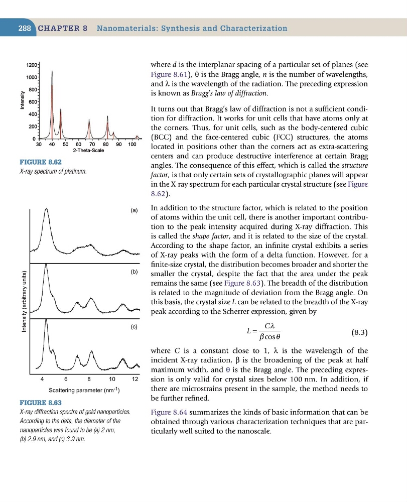
in the X-ray spectrum for each particular crystal structure (see Figure
8.62).
In addition to the structure factor, which is related to the position
of atoms within the unit cell, there is another important contribution to the peak intensity acquired during X-ray diffraction. This
is called the shape factor, and it is related to the size of the crystal.
According to the shape factor, an infinite crystal exhibits a series
of X-ray peaks with the form of a delta function. However, for a
finite-size crystal, the distribution becomes broader and shorter the
smaller the crystal, despite the fact that the area under the peak
remains the same (see Figure 8.63). The breadth of the distribution
is related to the magnitude of deviation from the Bragg angle. On
this basis, the crystal size L can be related to the breadth of the X-ray
peak according to the Scherrer expression, given by
L
C
=
λ
β
θ
cos
(8.3)
where C is a constant close to 1, λ is the wavelength of the
incident X-ray radiation, β is the broadening of the peak at half
maximum width, and θ is the Bragg angle. The preceding expression is only valid for crystal sizes below 100 nm. In addition, if
there are microstrains present in the sample, the method needs to
be further refined.
Figure 8.64 summarizes the kinds of basic information that can be
obtained through various characterization techniques that are particularly well suited to the nanoscale.
Figure 8.62
X-ray spectrum of platinum.
Figure 8.63
X-ray diffraction spectra of gold nanoparticles.
According to the data, the diameter of the
nanoparticles was found to be (a) 2 nm,
(b) 2.9 nm, and (c) 3.9 nm.
Intensity (arbitrary units)
Scattering parameter (nm -1 )
4
6
8
(a)
(b)
(c)
10
12
288
where d is the interplanar spacing of a particular set of planes (see
Figure 8.61), θ is the Bragg angle, n is the number of wavelengths,
and λ is the wavelength of the radiation. The preceding expression
is known as Bragg’s law of diffraction.
It turns out that Bragg’s law of diffraction is not a sufficient condition for diffraction. It works for unit cells that have atoms only at
the corners. Thus, for unit cells, such as the body-centered cubic
(BCC) and the face-centered cubic (FCC) structures, the atoms
located in positions other than the corners act as extra-scattering
centers and can produce destructive interference at certain Bragg
angles. The consequence of this effect, which is called the structure
factor, is that only certain sets of crystallographic planes will appear
in the X-ray spectrum for each particular crystal structure (see Figure
8.62).
In addition to the structure factor, which is related to the position
of atoms within the unit cell, there is another important contribution to the peak intensity acquired during X-ray diffraction. This
is called the shape factor, and it is related to the size of the crystal.
According to the shape factor, an infinite crystal exhibits a series
of X-ray peaks with the form of a delta function. However, for a
finite-size crystal, the distribution becomes broader and shorter the
smaller the crystal, despite the fact that the area under the peak
remains the same (see Figure 8.63). The breadth of the distribution
is related to the magnitude of deviation from the Bragg angle. On
this basis, the crystal size L can be related to the breadth of the X-ray
peak according to the Scherrer expression, given by
L
C
=
λ
β
θ
cos
(8.3)
where C is a constant close to 1, λ is the wavelength of the
incident X-ray radiation, β is the broadening of the peak at half
maximum width, and θ is the Bragg angle. The preceding expression is only valid for crystal sizes below 100 nm. In addition, if
there are microstrains present in the sample, the method needs to
be further refined.
Figure 8.64 summarizes the kinds of basic information that can be
obtained through various characterization techniques that are particularly well suited to the nanoscale.
Figure 8.62
X-ray spectrum of platinum.
Figure 8.63
X-ray diffraction spectra of gold nanoparticles.
According to the data, the diameter of the
nanoparticles was found to be (a) 2 nm,
(b) 2.9 nm, and (c) 3.9 nm.
Intensity (arbitrary units)
Scattering parameter (nm -1 )
4
6
8
(a)
(b)
(c)
10
12