

283
to large changes in tunneling current, giving STM the ability to
monitor very small variations in topography. In fact, if the system
is free from any kind of mechanical vibrations, an STM image has
sub-angstrom vertical resolution and atomic resolution laterally.
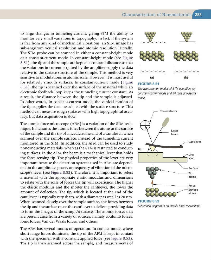
The STM probe can be scanned in either a constant-height mode
or a constant-current mode. In constant-height mode (see Figure
8.51), the tip and the sample are kept at a constant distance so that
the variations in current acquired by the amplifier supply the data
relative to the surface structure of the sample. This method is very
sensitive to modulations in atomic scale. However, it is most useful
for relatively smooth surfaces. In constant-current mode (Figure
8.51), the tip is scanned over the surface of the material while an
electronic feedback loop keeps the tunneling current constant. As
a result, the distance between the tip and the sample is adjusted.
In other words, in constant-current mode, the vertical motion of
the tip supplies the data associated with the surface structure. This
method can measure rough surfaces with high topographical accuracy, but data acquisition is slow.
The atomic force microscope (AFM) is a variation of the STM technique. It measures the atomic force between the atoms at the surface
of the sample and the tip of a needle at the end of a cantilever, when
scanned over the sample surface, instead of the tunneling current
monitored in the STM. In addition, the AFM can be used to study
nonconducting materials, whereas the STM is restricted to conducting surfaces. In the AFM, the beam is a mechanical lever that holds
the force-sensing tip. The physical properties of the lever are very
important because the detection systems used in AFM are dependent on the amplitude, phase, or frequency of vibration of the microscope’s lever (see Figure 8.52). Therefore, it is important to select
a material with the appropriate elastic modulus and dimensions
to relate with the scale of forces the tip will experience. The higher
the elastic modulus and the shorter the cantilever, the lower the
amount of deflection. The tip, which is located at the end of the
cantilever, is typically very sharp, with a diameter as small as 20 nm.
When scanned closely over the sample surface, the forces between
the tip and the surface cause the cantilever to deflect, providing data
to form the images of the sample’s surface. The atomic forces that
are present arise from a variety of sources, namely coulomb forces,
ionic forces, Van der Waals forces, and others.
The AFM has several modes of operation. In contact mode, where
short-range forces dominate, the tip of the AFM is kept in contact
with the specimen with a constant applied force (see Figure 8.53).
The tip is then scanned across the sample, and measurements of
Characterization of Nanomaterials
(a)
(b)
Figure 8.51
The two common modes of STM operation: (a)
constant-current mode and (b) constant-height
mode.
Figure 8.52
Schematic diagram of an atomic force microscope.
Tip
atoms
Surface
Line
scan
Tip
Cantilever
Photodetector
Laser
beam
Surface
atoms
Force
to large changes in tunneling current, giving STM the ability to
monitor very small variations in topography. In fact, if the system
is free from any kind of mechanical vibrations, an STM image has
sub-angstrom vertical resolution and atomic resolution laterally.
The STM probe can be scanned in either a constant-height mode
or a constant-current mode. In constant-height mode (see Figure
8.51), the tip and the sample are kept at a constant distance so that
the variations in current acquired by the amplifier supply the data
relative to the surface structure of the sample. This method is very
sensitive to modulations in atomic scale. However, it is most useful
for relatively smooth surfaces. In constant-current mode (Figure
8.51), the tip is scanned over the surface of the material while an
electronic feedback loop keeps the tunneling current constant. As
a result, the distance between the tip and the sample is adjusted.
In other words, in constant-current mode, the vertical motion of
the tip supplies the data associated with the surface structure. This
method can measure rough surfaces with high topographical accuracy, but data acquisition is slow.
The atomic force microscope (AFM) is a variation of the STM technique. It measures the atomic force between the atoms at the surface
of the sample and the tip of a needle at the end of a cantilever, when
scanned over the sample surface, instead of the tunneling current
monitored in the STM. In addition, the AFM can be used to study
nonconducting materials, whereas the STM is restricted to conducting surfaces. In the AFM, the beam is a mechanical lever that holds
the force-sensing tip. The physical properties of the lever are very
important because the detection systems used in AFM are dependent on the amplitude, phase, or frequency of vibration of the microscope’s lever (see Figure 8.52). Therefore, it is important to select
a material with the appropriate elastic modulus and dimensions
to relate with the scale of forces the tip will experience. The higher
the elastic modulus and the shorter the cantilever, the lower the
amount of deflection. The tip, which is located at the end of the
cantilever, is typically very sharp, with a diameter as small as 20 nm.
When scanned closely over the sample surface, the forces between
the tip and the surface cause the cantilever to deflect, providing data
to form the images of the sample’s surface. The atomic forces that
are present arise from a variety of sources, namely coulomb forces,
ionic forces, Van der Waals forces, and others.
The AFM has several modes of operation. In contact mode, where
short-range forces dominate, the tip of the AFM is kept in contact
with the specimen with a constant applied force (see Figure 8.53).
The tip is then scanned across the sample, and measurements of
Characterization of Nanomaterials
(a)
(b)
Figure 8.51
The two common modes of STM operation: (a)
constant-current mode and (b) constant-height
mode.
Figure 8.52
Schematic diagram of an atomic force microscope.
Tip
atoms
Surface
Line
scan
Tip
Cantilever
Photodetector
Laser
beam
Surface
atoms
Force