

277
Characterization of Nanomaterials
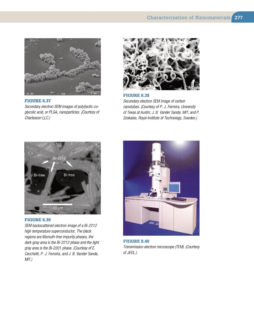
Figure 8.37
Secondary electron SEM images of poly(lactic-coglycolic acid, or PLGA, nanoparticles. (Courtesy of
Charlesson LLC.)
Figure 8.38
Secondary electron SEM image of carbon
nanotubes. (Courtesy of P. J. Ferreira, University
of Texas at Austin; J. B. Vander Sande, MIT; and P.
Szakalos, Royal Institute of Technology, Sweden.)
10 µm
Bi-free
Bi-free
Bi-2212
Figure 8.39
SEM backscattered electron image of a Bi-2212
high temperature superconductor. The black
regions are Bismuth-free impurity phases, the
dark-gray area is the Bi-2212 phase and the light
gray area is the Bi-2201 phase. (Courtesy of E.
Cecchetti, P. J. Ferreira, and J. B. Vander Sande,
MIT.)
Figure 8.40
Transmission electron microscope (TEM). (Courtesy
of JEOL.)
Characterization of Nanomaterials
Figure 8.37
Secondary electron SEM images of poly(lactic-coglycolic acid, or PLGA, nanoparticles. (Courtesy of
Charlesson LLC.)
Figure 8.38
Secondary electron SEM image of carbon
nanotubes. (Courtesy of P. J. Ferreira, University
of Texas at Austin; J. B. Vander Sande, MIT; and P.
Szakalos, Royal Institute of Technology, Sweden.)
10 µm
Bi-free
Bi-free
Bi-2212
Figure 8.39
SEM backscattered electron image of a Bi-2212
high temperature superconductor. The black
regions are Bismuth-free impurity phases, the
dark-gray area is the Bi-2212 phase and the light
gray area is the Bi-2201 phase. (Courtesy of E.
Cecchetti, P. J. Ferreira, and J. B. Vander Sande,
MIT.)
Figure 8.40
Transmission electron microscope (TEM). (Courtesy
of JEOL.)